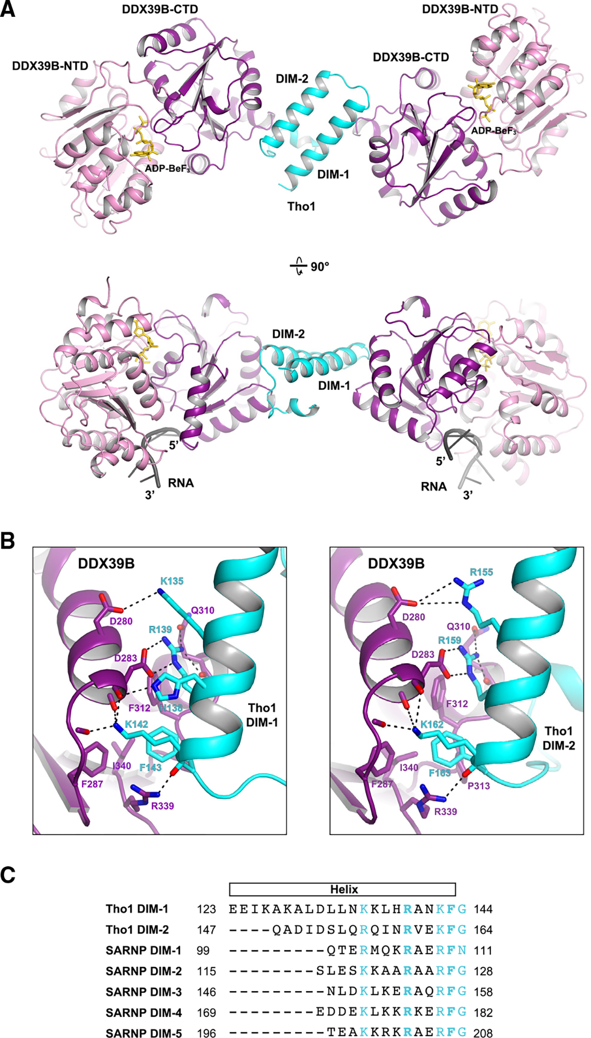
Figure 2. Crystal structure of a Tho1/DDX39B/RNA complex at 2.5 Å resolution.

(A) DDX39B was crystallized with a C-terminal domain of yeast Tho1 in the presence of poly(U) 15-mer RNA and the non-hydrolyzable ATP analog ADP-BeF3. The model shown in two orientations features a 1:2 assembly of Tho1 and DDX39B. The structure reveals a DDX39B interacting motif (DIM) that recognizes the DDX39B-CTD domain.
(B) DDX39B interfaces with DIM-1 (left) and DIM-2 (right) of Tho1.
(C) Alignment of the two DIMs in yeast Tho1 and five DIMs in human SARNP. Conserved residues are highlighted in cyan. The two invariant residues (R5 and F9) are shown in bold.
