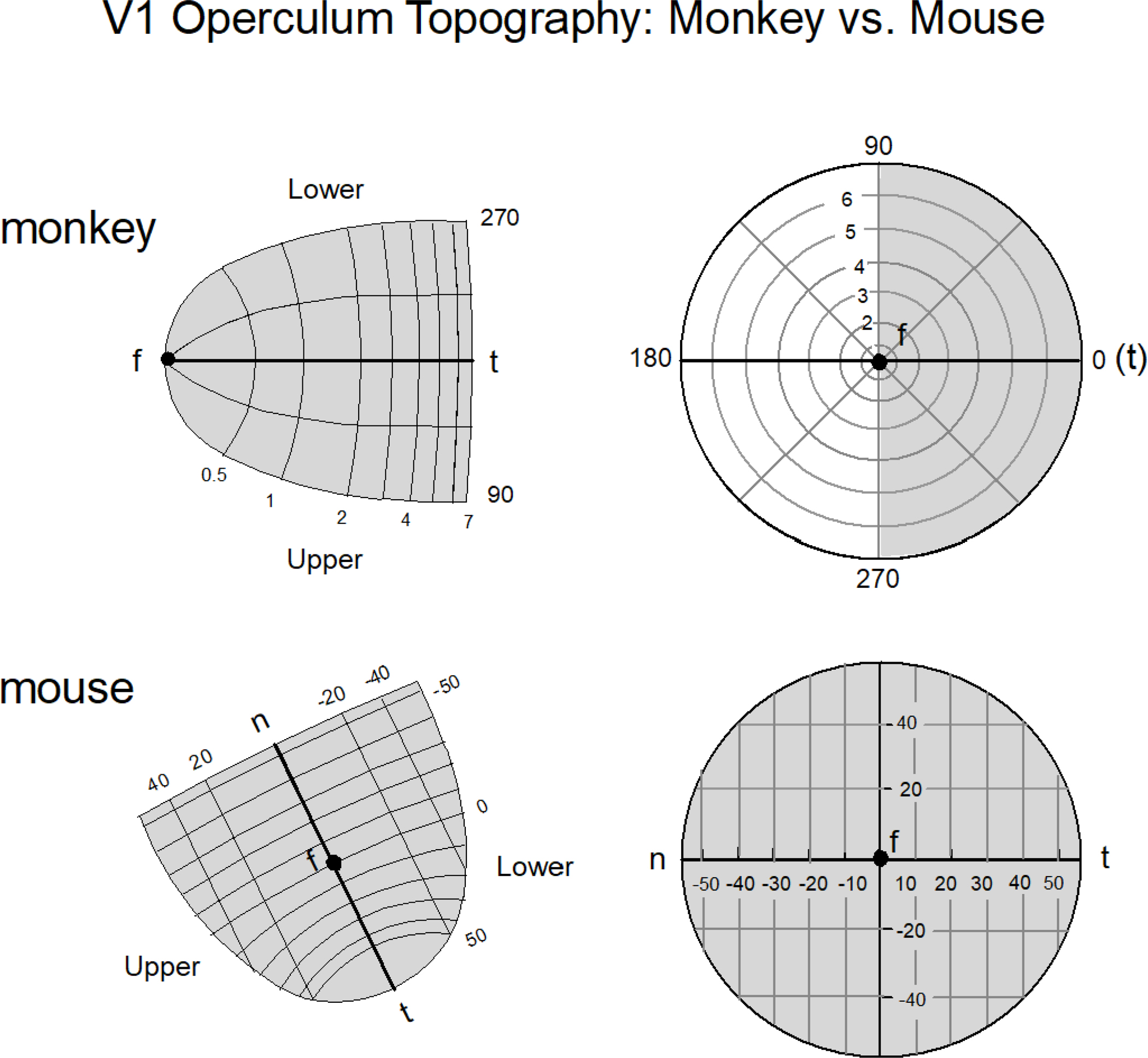
Figure 2.

The layout of area V1 of the macaque monkey and the mouse as it pertains to the operculum (i.e., the exposed area of neocortex) is shown. In the case of the macaque monkey only 7 degrees of the visual field of one hemifield is represented in the operculum (top panel)(derived from Schiller and Tehovnik 2008); in the case of the mouse the arrangement is different (bottom panel): the entire visual field is encoded by the operculum and the center of gaze marked by ‘f’ is situated in the center of the map with ‘n’ representing the nasal field and ‘t’ representing temporal field (derived from Fig. 1G,H & Fig. 7A,D of Garret et al. 2014). One operculum in the mouse encodes the entire visual field from a viewing eye as illustrated. Notice the slight magnification of the visual representation beyond the center of gaze ‘f’ for the nasal representation of the mouse; for the macaque monkey the magnification is more extreme which accounts for its superior visual spatial resolution. The magnification is a rough approximation. For precise depictions see Schiller and Tehovnik (2008) and Garret et al. (2014).
