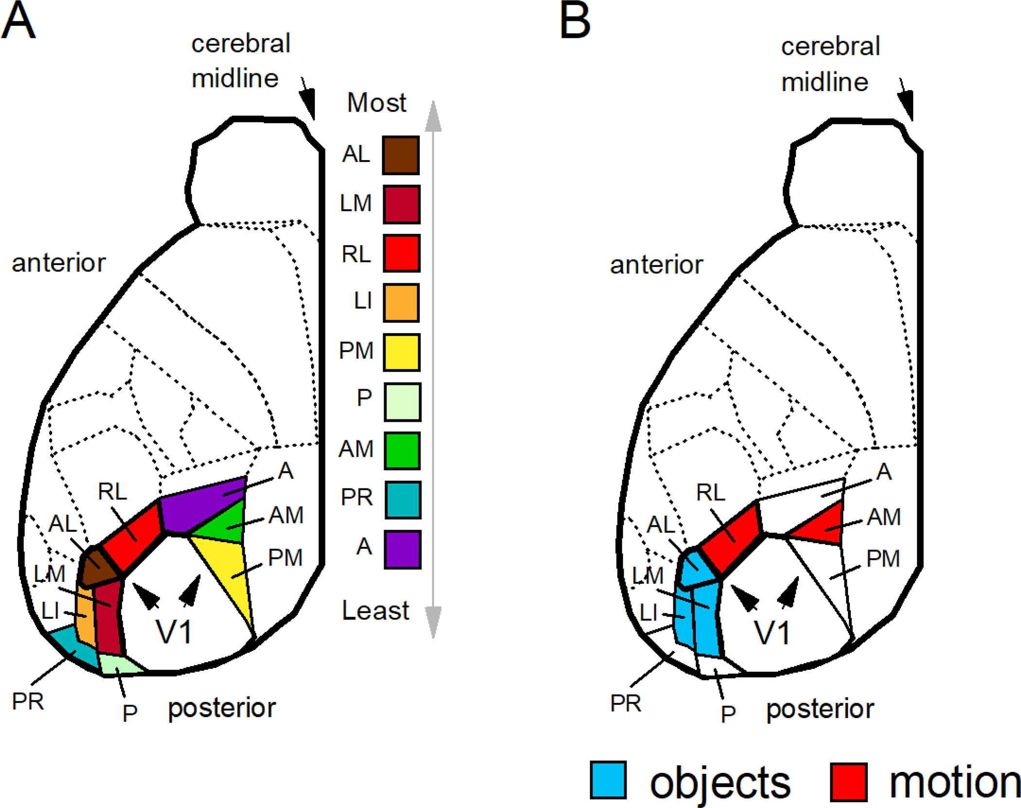
Figure 4.

(A) Extrastriate areas of the mouse are listed from top to bottom according to the density of innervation from V1 from maximal to minimal as derived from Froudarakis et al. (2019): the anterolateral area (AL), the lateromedial area (LM), the rostrolateral area (RL), the lateral intermediate area (LI), the posteromedial area (PM), the posterior area (P), the anteromedial area (AM), the postrhinal area (PR), and the anterior area (A). (B) Neurons that are modulated by objects have been identified in the anterolateral (AL), lateromedial (LM), and lateral intermediate (LI) areas (defined in blue); neurons modulated by complex motion stimuli (e.g. flow fields) have be identified in the rostrolateral (RL) and anteromedial (AM) areas (defined in red). Regions of the extrastriate cortex that encode objects are located lateral to the regions that encode motion. Complete details of the mouse visual cortex can be found in Froudarakis et al. (2019, 2020), Garrett et al. (2014), Marshel et al. (2011), Rasmussen et al. (2020), and Wang et al. (2012).
