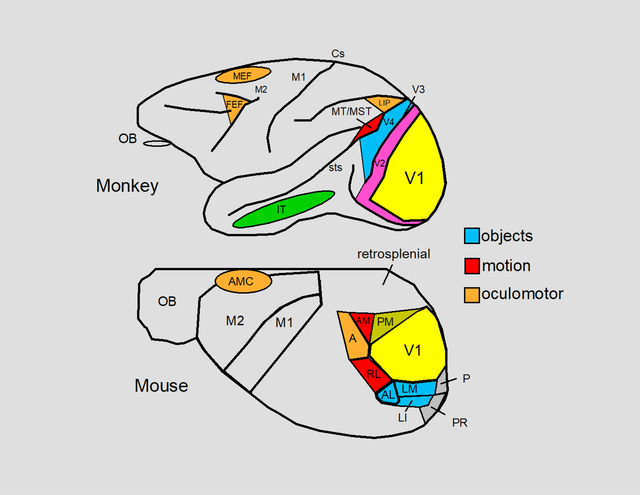
Figure 5.

The visual areas of the neocortex of the macaque monkey and the mouse are summarized. Both species have homologous areas for processing visual information starting at V1 which process stationary and moving oriented lines. Object encoding has been described for V4 in the macaque monkey and for areas AL (anterolateral), LM (lateromedial) and LI (lateral intermediate) in the mouse. V4 of the macaque monkey ultimately innervates the IT (inferior temporal) cortex which contains cells that respond to faces and other complex objects. The object encoding areas of both the mouse and the macaque monkey contain a central-field representation. Motion encoding has been described for MT/MST (middle temporal cortex/middle superior temporal cortex) in the macaque monkey and for areas AM (anteromedial) and RL (rostrolateral) in the mouse. MT and MST ultimately innervate LIP (the lateral interparietal area) which is an oculomotor area that mediates eye movements and active fixation in macaque monkeys (Andersen and Mountcastle 1983; Mountcastle et al. 1975). Area A (anterior) of the mouse may be a homologue of LIP for eye movements can be evoked from this region (Itokazu et al. 2018) and this area has been implicated in spatial vision in rats (Kolb and Walkey 1987). In the macaque monkey, the visual signals of the posterior cortex eventually arrive in the frontal lobes at one of the two major oculomotor areas: the FEF (the frontal eye fields) and the MEF (the medial eye fields). The FEF is a central controller of eye movements (saccadic, smooth pursuit, and vergence) and the MEF is involved in eye, head, and body part coordination. Activation of the AMC (anteromedial cortex) in the mouse evokes eye movements (Itokazu et al. 2018) as well as head movements in rodents such as rats (Tehovnik and Yeomans 1987). Whether the AMC contains FEF and MEF homologues is not known. V2, V3, sts (superior temporal sulcus), and Cs (central sulcus) are indicated for the macaque monkey and areas PM (posteromedial), P (posterior), and PR (postrhinal) are indicated for the mouse. The remaining labels include M1, M2, the retrosplenial cortex, and the olfactory bulb (OB). The inset to the right color codes some of the areas according to function: objects (blue), motion (red), and oculomotor (orange). For further details see: Froudarakis et al. (2019), Garrett et al. (2014), Marshel et al. (2011), Rasmussen et al. (2020), and Schiller and Tehovnik (2015).
