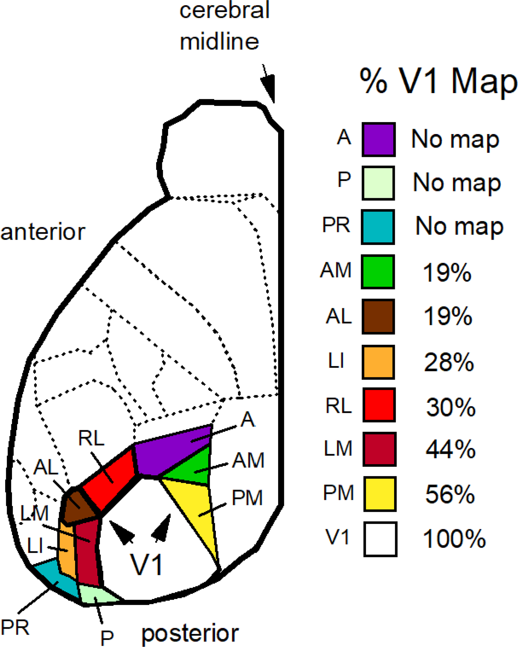
Figure 6.

Extrastriate areas of the mouse are listed from top to bottom according to the percent of the visual field represented as a fraction of that represented by the V1 map using the data of figures 5B,C of Garrett et al. (2014). All maps contained a central visual field encoding the primary optical axis. Regions are list from no topographic coverage (no map) to maximal coverage: the anterior area (A), the posterior area (P), postrhinal area (PR), the anteromedial area (AM), the anterolateral area (AL), the lateral intermediate area (LI), the rostrolateral area (RL), the lateromedial area (LM), the posteromedial area (PM), and area V1 (V1).
