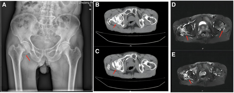
Figure 1.
(A) Pelvic X-ray: An oblique translucent line was seen under the right femoral head, and the broken end was slightly separated and incarcerated. (B and C) Bilateral hip CT: the cortex of the right femoral neck was discontinuous, and the broken end was slightly angled and incarcerated. There was no obvious displaced fracture of the left femoral neck. (D and E) Right hip MRI: discontinuous cortical bone of the right femoral neck, edema of the surrounding bone marrow, displacement of the broken end, and swelling of the surrounding soft tissue. Left femoral neck edema with abnormal signal can be seen. CT = computed tomography.

