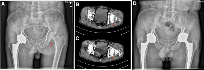
Figure 3.
(A) Pelvic X-ray: left femoral neck fracture with dislocation and displacement. Good prosthesis position after right THA. (B and C) Bilateral hip CT: Displaced fracture of the left femoral neck with osteosclerosis of the broken end. (D) Postoperative pelvic X-ray: good prosthesis position after bilateral hip THA. CT = computed tomography, THA= total hip arthroplasty.

