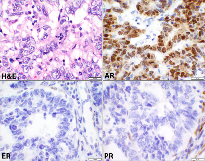
Fig 2. Androgen, estrogen, and progesterone receptors in serous carcinoma.
Hematoxylin and eosin (H&E) stain of a case of uterine serous carcinoma showing papillary and glandular patterns where cells exhibit high-grade cytology, mitotic figure, and marked nuclear pleomorphism (case #40, S1 Table). The corresponding androgen receptor (AR), estrogen receptor (ER), and progesterone receptor (PR) immunostains show positive nuclear staining with AR and negative nuclear staining of the malignant cells for ER and PR (40× objective).

