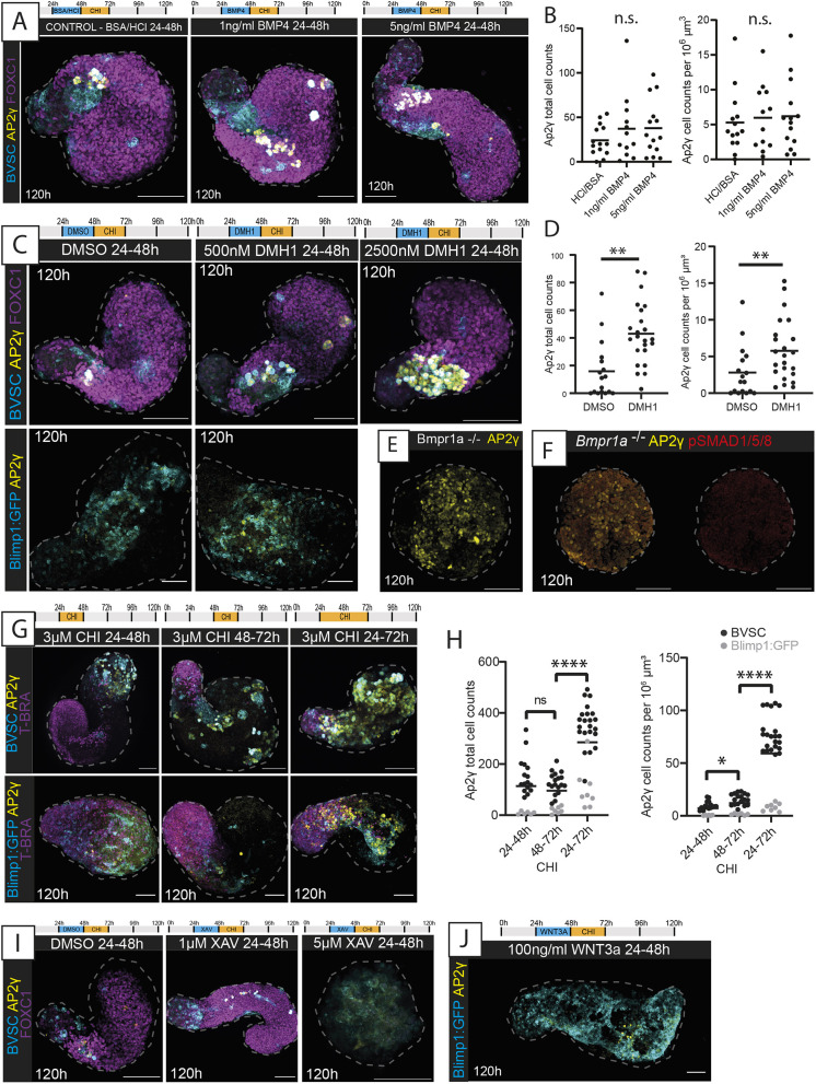
Fig. 6.
BMP and Wnt signalling modulation in Gld-PGCLCs. (A) Maximum-projection images of BVSC gastruloids following BMP4 application at the timepoints and concentrations indicated. In BVSC gastruloids, Blimp1:mVenus is membrane-targeted, whereas Stella:eCFP is found throughout the cell. (B) Quantification of AP2γ+ cells in the conditions indicated from BVSC and Blimp1:GFP gastruloids at 120 h. (C) Maximum-projection images of gastruloids following BMP inhibition by DMH1 application at the timepoints and concentrations indicated. (D) Quantification of AP2γ+ cells following DMSO or 500 nM DMH1 treatment, from BVSC and Blimp1:GFP gastruloids at 120 h. (E) Gastruloids made from the Bmpr1a−/− cell line, showing aberrant morphology with lack of elongation and significant numbers of AP2γ+ cells. (F) Absence of pSMAD1/5/8 in Bmpr1a−/− gastruloids at 120 h. (G) Maximum-projection images of gastruloids exposed to different timings of CHIR-99021 (CHI) application as indicated. (H) Quantification of AP2γ+ cells in the conditions indicated, from BVSC and Blimp1:GFP gastruloids at 120 h. (I) Maximum-projection images of BVSC gastruloids exposed to XAV939 (XAV) to inhibit Wnt signalling. (J) Maximum-projection image of a Blimp1:GFP gastruloid exposed to WNT3A at the timepoint shown. Black bars (B,D,H) represent the mean average. Dashed lines, morphological gastruloid outlines from Hoechst staining. Images are representative of 4-22 gastruloids per panel. Scale bars: 100 µm. n.s., no significant differences; *P<0.05; **P<0.01; ****P<0.0001 (unpaired, two-tailed t-test with Welch's correction).

