Fig. 2.
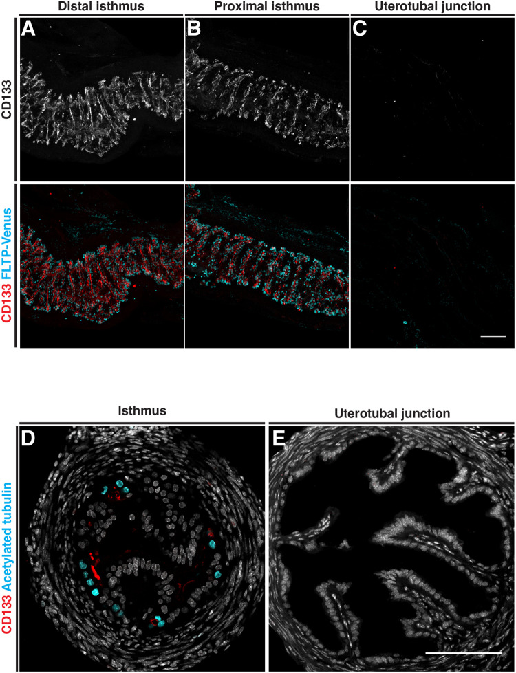
Distribution of CD133 in the proximal adult mouse oviduct. The distribution patten of CD133 analyzed by immunostaining in FLTP-H2B-Venus mice. (A,B) Whole-mount Z-projection of the distal and proximal isthmus showing CD133 staining along transverse epithelial folds associated with multiciliated cells labeled with FLTP-Venus. (C) Whole-mount Z-projection of the uterotubal junction show no CD133 staining or FLTP-Venus cells. (D) A transverse section of the isthmus showing CD133 on the apical surface of FLTP-Venus multiciliated epithelial cells located at the base of epithelial folds. (E) A transverse section of the uterotubal junction confirming the absence of CD133 and FLTP-Venus cells. Scale bars: 100 µm.

