Fig. 4.
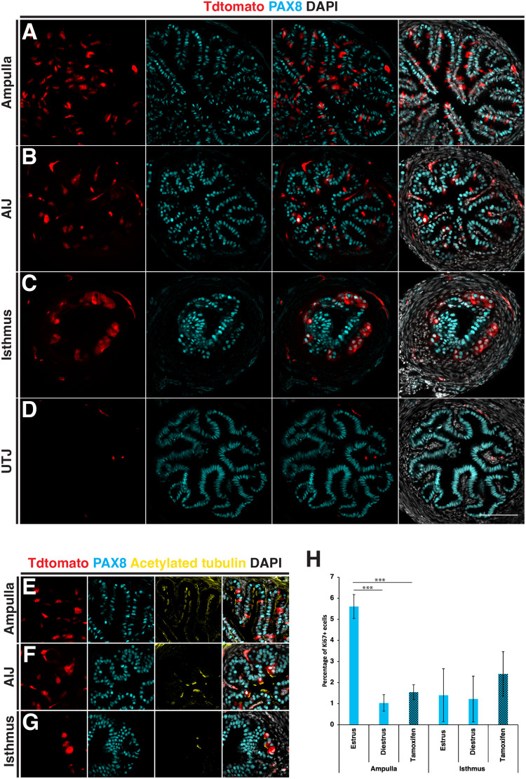
Distribution of Prom1-expressing cells in the adult mouse oviduct. The distribution pattern of Prom1 expressing cells was analysed in Prom1C-L:C-L:Rosa26Tdtomato:Tdtomato mice, by tamoxifen injection on 5 consecutive days to label Prom1-expressing cells and dissection of oviducts after 72 h. (A) Transverse section of the ampulla showing a diffuse pattern of Tdtomato-labeled cells counter stained with a PAX8 antibody to label secretory cells. (B) Fewer Tdtomato-labeled cells were detected in the ampulla–isthmus junction. (C) Tdtomato-labeled cells were restricted to the base of epithelial folds in the isthmus. (D) No Tdtomato-labeled cells were identified in the epithelial cells at the uterotubal junction. (E-G) Counter staining with cilia marker acetylated tubulin, revealed the majority of Tdtomato cells to be multiciliated in the ampulla, ampulla–isthmus junction and isthmus. (H) A comparison of the proportion of Ki67- positive epithelial cells during the oestrus cycle and 6 h after tamoxifen administration. No significant changes were detected in the isthmus, while significantly more Ki67-positive cells were detected during estrus compared to diestrus and after tamoxifen administration. Statistical tests used in H=two-tailed Student's t-test. Scale bars: A-D, 100 µm; E-G, 10 µm.

