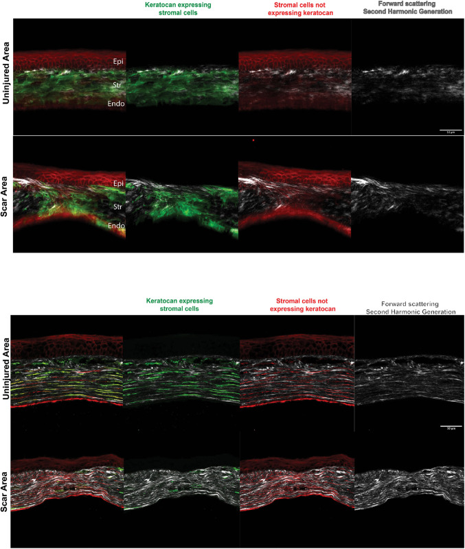
Fig. 3.
Imaging of mature corneal scars using SHG and fluorescence microscopy in a freshly enucleated adult I-KeramTmG eye mounted for imaging shows keratocytes in the repairing stroma 3 months after injury. A doxycycline diet was given 1 week prior to injury to assess eGFP expression in scars. Top: uninjured corneas had a predominance of keratocytes embedded within the corneal stroma, with few cells expressing tdTomato. Second-harmonic generation (SHG) imaging (gray, right) showed horizontally well-aligned and organized collagen fibrils/lamellae in the stroma. The scar area showed a mixture of keratocytes and tdTomato-expressing cells in the scar tissue, which was disorganized in an oblique and vertical array. Similarly, collagen fibrils/lamellae were disorganized (n=4). Bottom: tissue sections showed keratocytes in the repairing stroma 3 months after injury. Uninjured corneas had a predominance of keratocytes embedded within the corneal stroma (green and red signals), with a minimal number of cells expressing tdTomato (red). Cells were parallel to the basement and Descemet's membranes. SHG imaging (gray, right) showed well-aligned and horizontally organized collagen stromal fibrils/lamellae. Scars had a mixture of keratocytes and tdTomato-expressing cells oriented in a disorganized fashion. Similarly, collagen fibrils/lamellae were disorganized (n=6). Epi, epithelium; Str, stroma; Endo, endothelium. Scale bars: 50 µm.

