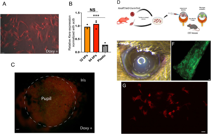
Fig. 4.
Keratocan expression is lost during standard in vitro expansion in plastic culture dishes with serum stimulation but maintained or reacquired if cells are in their unique stromal niche. (A) Freshly passed fibroblasts expanded in vitro under serum stimulation shut down keratocan expression, despite maintaining a dendritic morphology (n=3). (B) Stromal cells downregulated keratocan expression when cultured in a stiffer matrix; plastic dishes in this case (n=3). In contrast, keratocytes expressed eGFP ex vivo – in serum medium supplemented with doxycycline – when maintained within the stromal matrix. NS, not significant; ***P=0.0003 (two-tailed unpaired t-test). (C) The image shows induced cells (eGFP expression) that are more noticeable in the region of the pupil 24 h after exposure to doxycycline in the culture medium (n=5). (D) Expanded tenocytes – known to express keratocan in vivo – or corneal-derived fibroblasts can be expanded in vitro and transplanted to the stromal matrix to evaluate whether keratocan is expressed. (E) An area of corneal edema (dashed circle) developed immediately after cell injection; the edema resolved in a few days and corneal transparency returned to normal (n=7). (F) eGFP expression in tendons in vivo after 1 week of oral doxycycline diet (n=6). (G) However, expanded tenocytes failed to express keratocan (eGFP) when transplanted to the stromal matrix (n=5). Scale bars: 25 µm (A,C,F,G).

