Fig. 4.
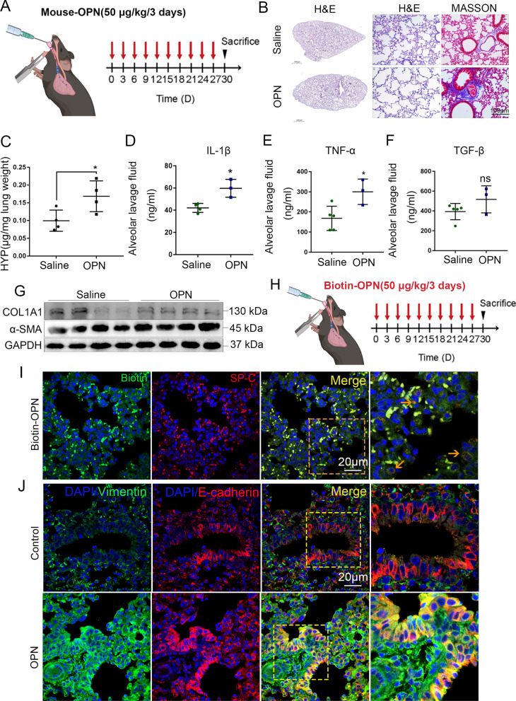
OPN effects alveolar epithelial cell and promotes lung fibrogenesis and the development of PF. A Schematic overview of experimental design for (B–G), recombinant mouse OPN in 50 µl of PBS (50 mg/kg) was instilled into the trachea every 3 days for one month. (n≥4). B H&E and Masson staining of lung tissues after multiple OPN exposures. Scale bar, 1000 μm or 100 μm. C Lung tissue hydroxyproline content was measured in differently treated animals. *P < 0.05; means + SD. D–F Concentration of IL-1β (D), TNF-α (E) and TGF-β (F) in bronchoalveolar lavage fluid (BALF) determined by ELISA. *P < 0.05, ns denotes no significance compared with saline group; means + SD. G Immunoblots of collagen and α-SMA of mouse lung tissue. H Schematic overview of experimental design for (I, J), recombinant mouse biotin-OPN in 50 µl of PBS (50 mg/kg) was instilled into the trachea every 3 days for one month. (n≥4). I IF of streptavidin-Texas (red) with SP-C (green) in biotin-OPN challenged mice. Scale bars, 20 μm. The orange arrows indicate immunofluorescence co-locatable cells. J Confocal microscopy showing vimentin (green), E-cadherin (red), and DNA dye DAPI (cyan) in lung tissue section of mice treated with OPN and matched group, showing epithelial-mesenchymal transition. Bars, 20 µm

