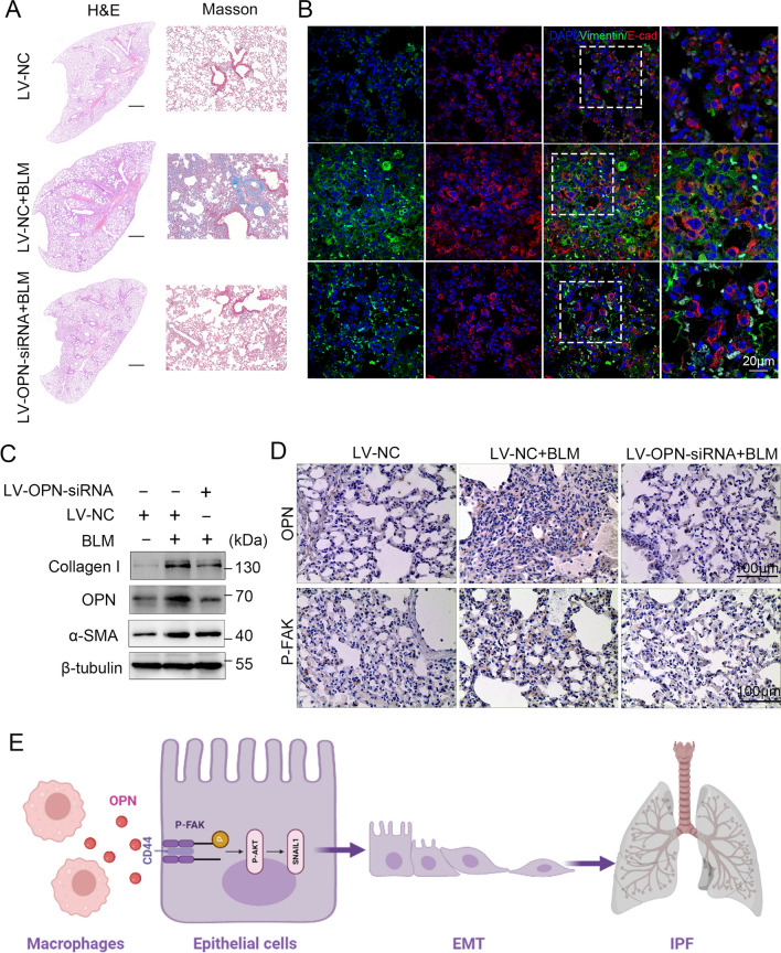
Fig. 7.
Silence of OPN attenuates epithelial-mesenchymal transition and pulmonary fibrosis. Mice (n = 10 in each group) were intratracheally injected with 1 × 10⁹ TU LV-OPN-siRNA or negative control (NC) 7 days after administration of bleomycin. Mice were killed at day 21 after BLM instillation. A Pulmonary fibrosis was determined by hematoxylin–eosin (H&E) staining and Masson’s trichrome staining. Representative images of three independent experiments are shown. B Representative IHC OPN and P-FAK staining of mouse lung tissue. C Immunoblots of OPN, collagen and α-SMA of mouse lung tissue. D Confocal microscopy showing vimentin (green), E-cadherin (red), and DNA dye DAPI (cyan) in lung tissue section of mice, showing epithelial-mesenchymal transition. Bars, 100 µm. E Summary of the article

