Fig. 3.
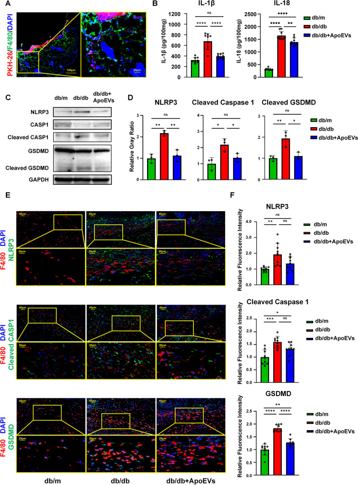
UCMSC-derived ApoEVs inhibited macrophage pyroptosis present during skin defect healing in db/db mice. a Immunofluorescence staining shows UCMSC-derived ApoEVs phagocytosed by F4/80-positive macrophages in vivo. Scale bar, 100 μm in low-magnification images, 20 μm in high-magnification images. b Tissue inflammatory factors IL-1β and IL-18 of different groups released in and around mouse skin defect area detected by ELISA, N = 3 per group, 3 duplication per sample. c Tissue protein levels of NLRP3, caspase-1, cleaved caspase-1, gasdermin D, cleaved gasdermin D of different groups released in and around mouse skin defect area detected by Western Blots, N = 3 per group. Corresponding uncropped full-length gels and blots are presented in Additional file 5B. d Semi-quantification of protein expression of NLRP3, cleaved caspase-1, and cleaved gasdermin D. e Immunofluorescence staining shows NLRP3, cleaved caspase-1, and gasdermin D in the skin defect area of mice in all three groups had significant co-localization with F4/80-positive macrophages. Scale bar, 40 μm in low-magnification images, 20 μm in high-magnification images. N = 3 per group, 3 ROIs per sample. f Semi-quantification of NLRP3, cleaved caspase-1, and gasdermin D relative fluorescence intensity in F4/80-positive cells of skin defect area. Data shown as mean ± SD. *P < 0.05; **P < 0.01; ***P < 0.001; ****P < 0.0001; NS not significant. CASP1 caspase-1; GSDMD, gasdermin D

