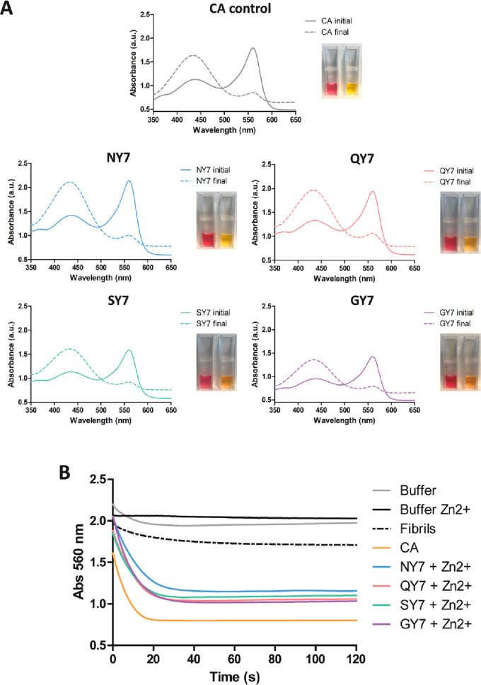
Figure 6.
Carbonic anhydrase activity of the fibrils in the presence of Zn2+. (A) Carbonic anhydrase (CA) activity was detected by measuring absorbance of the phenol red pH indicator in the presence of NY7 (blue), QY7 (red), SY7 (green), and GY7 (purple) and positive control with CA at 50 nM (gray). Initial (continuous line) and final (discontinuous line) spectra were acquired at between 350 and 650 nm. The image shows the phenol red coloration corresponding to the initial (red) and final (yellow) samples. (B) Time-dependent decrease in absorbance at 560 nm of phenol red incubated with CO2-treated deionized water. Fibril colors as in (A), negative controls of buffer alone (gray), buffer with Zn2+ (black), QY7 fibrils without metal (black discontinuous), and CA at 50 nM (orange) are indicated.

