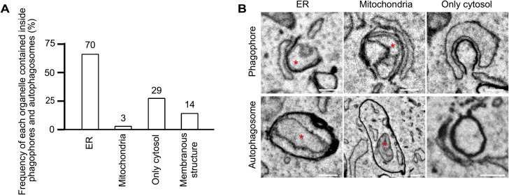
Fig. 5.
Autophagosomal contents in phagophores and autophagosomes
(A) From 3D-EM images of 28 phagophores and 79 autophagosomes (total 107), the organelles inside were defined based on morphology. ER fragments were identified as ribosome-bound organelles, tubular structures with a single membrane having less electron density compared with autophagosomal membranes, or organelles connected to the ER outside. Mitochondria were identified as organelles with cristae. “Membranous structures” indicate organelles not specified as either the ER or mitochondria. (B) The representative EM images of phagophores and autophagosomes containing indicated organelles (red asterisks). Scale bar, 0.2 μm.

