Fig. 4.
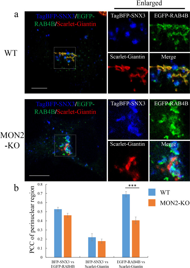
Localization of RE in WT and MON2-KO cells. a, Representative merged confocal images of RAB4B-positive RE. HEK293 WT or MON2-KO cells were transiently transfected with TagBFP-SNX3, EGFP-RAB4B, and Scarlet-Giantin. Enlarged images show magnified views of the boxed areas. Scale bars, 10 μm. b, Quantitative analysis of co-localization between TagBFP-SNX3, EGFP-RAB4B, and Scarlet-Giantin in the perinuclear region. PCCs between two proteins were calculated using the ImageJ plugin JACoP. The data represent means±SE of the measurements (N=12 for WT cells; N=10 for MON2-KO cells). ***, P<0.001 (two-tailed Student’s t test).

