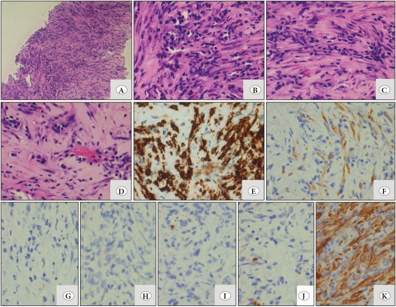
Figure 2.
A) Low magnification shows core biopsy with proliferation of bland spindle cells (H&E; x20). B-D) High magnification shows different areas observed in the biopsy such as areas with predominance of lymphoplasmacytic infiltrate and spindle cells arranged in long fascicles. (H&E; x40). E-F) IHC shows comparison of ALK immunostains, on both the Dako platform using monoclonal mouse anti-human CD246 antibody (clone ALK1) which showed focal positivity with weak intensity (F; x40) and the Ventana platform using monoclonal rabbit anti-human CD246 antibody (clone D5F3) reported as strong positive (E; x40). Spindle cells are negative for the following: G) Desmin H) CK I) CD117 and J) S-100 (IHC; x40). K) SMA positivity in neoplastic cells (IHC; x40).

