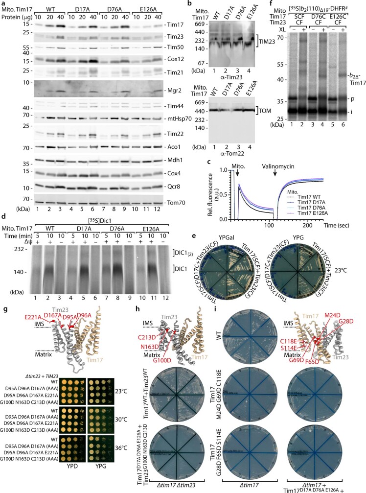
Extended Data Fig. 6. Tim17D17A and Tim17D76A mitochondria have no obvious secondary defects and introduction of equivalent negative charges into Tim23 or lower parts of the lateral cavity of Tim17.
a, Protein amounts of WT, Tim17D17A, Tim17D76A and Tim17E126A mitochondria isolated from yeast cells grown in YPG at 23 °C, followed by SDS-PAGE and immunodecoration with the indicated antibodies. b, Protein complexes of isolated Tim17 WT and mutant yeast mitochondria analysed by BN-PAGE and immunodecoration with the indicated antibodies. TIM23, presequence translocase of the inner membrane; TOM, translocase of the outer mitochondrial membrane. c, Assessment of the mitochondrial membrane potential of isolated mitochondria as described in Extended Data Fig. 2d. d, Import of radiolabelled metabolite carrier protein Dic1 into isolated WT and tim17 mutant mitochondria, followed by BN-PAGE and autoradiography. e, Growth analysis of tim17 tim23 mutant yeast strains expressing Tim17SCF+Tim23CF or Tim17D17C+Tim23CF on agar media containing galactose (YPGal) and glycerol (YPG) at 23 °C. f, Import of 35S-labelled b2(110)∆19-DHFR# into Tim17 WT or cysteine mutant mitochondria in the presence of MTX followed by chemical crosslinking (MBS; XL), SDS-PAGE and autoradiography. g, ColabFold structural protein complex model of S. c. Tim17 (tan) and Tim23 (grey) heterodimer with the location of the negatively charged residues (red) within the transmembrane domains and adjacent segments of Tim23 (upper panel). Growth analysis of tim23 mutant yeast strains expressing Tim23 WT or negative charge mutant variants as for Extended Data Fig. 2b (lower panel). AAA, Tim23 D95A D96A D167A. h, i, ColabFold structural protein complex model as for g with the location of residues equivalent to Tim17 D17, D76 and E126 that were introduced into the transmembrane regions of Tim23 (h) or locations with equivalent charged residues, located lower within the lateral cavity of Tim17, were introduced (i) (red; upper panels). Growth phenotypes of tim17 tim23 mutant yeast strains in the tim17∆tim23∆ background (h) or tim17 mutants in the tim17∆ and tim17∆, Tim17D17A_D76A_E126A background (i) on 5-FOA medium at 23 °C (lower panel).

