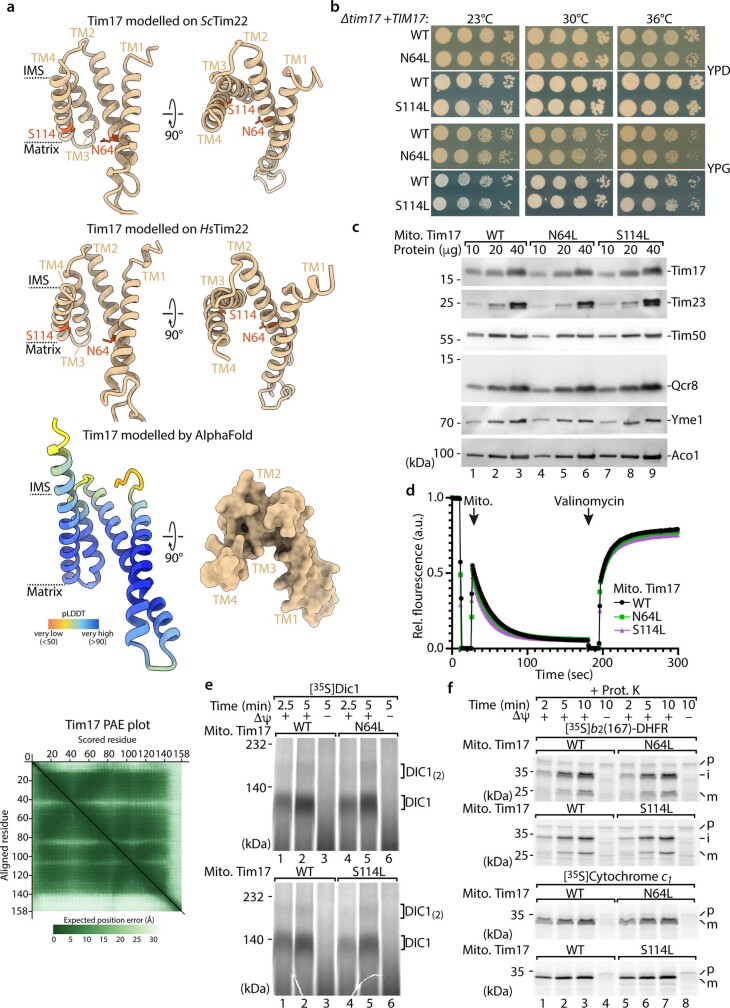
Extended Data Fig. 2. Hydrophilic residues within the lateral Tim17 cavity are crucial for matrix translocation.
a, Tim17 (S. cerevisiae) modelled on the Tim22 (S.c.) structure (top; PDB ID 6LO8) and Tim22 (H. sapiens) structure (middle; PDB ID 7CGP). Dashed lines indicate the transmembrane (TM) segments of Tim17 (orange, asparagine 64 (N64), and serine (S114) residues). Predicted Local Distance Difference Test (pLDDT) score together with predicted aligned error plot (PAE) as well as surface representation of Tim17 as seen from the intermembrane space of the Tim17 AlphaFold model depicted in Fig. 1e (bottom). IMS, intermembrane space. b, Growth analysis of yeast strains expressing WT, Tim17N64L or Tim17S114L variants on agar with fermentable (dextrose/glucose, YPD) and non-fermentable (glycerol, YPG) medium at the indicated temperatures. c, Protein amounts of WT, Tim17N64L and Tim17S114L mitochondria isolated from yeast cells grown on non-fermentable media (YPG) at 23 °C, analysed by SDS-PAGE and immunodecoration against the indicated antibodies. d, Membrane potential assessment of isolated WT, Tim17N64L and Tim17S114L mitochondria (from yeast cells grown in YPG at 23 °C). The membrane potential was assessed by fluorescence quenching using the potential sensitive dye 3,3’-dipropylthiadicarbocyanine iodide and subsequently dissipated by addition of valinomycin. e, Import of radiolabelled metabolite carrier protein dicarboxylate carrier 1 (Dic1) into isolated WT, Tim17N64L and Tim17S114L mitochondria, followed by blue native PAGE and autoradiography. Δψ, membrane potential; DIC1(2), assembled Dic(oligomer). f, Import of radiolabelled b2(167)-DHFR and cytochrome c1 into isolated WT, Tim17N64L and Tim17S114L mitochondria followed by SDS-PAGE and autoradiography.

