Fig. 2.
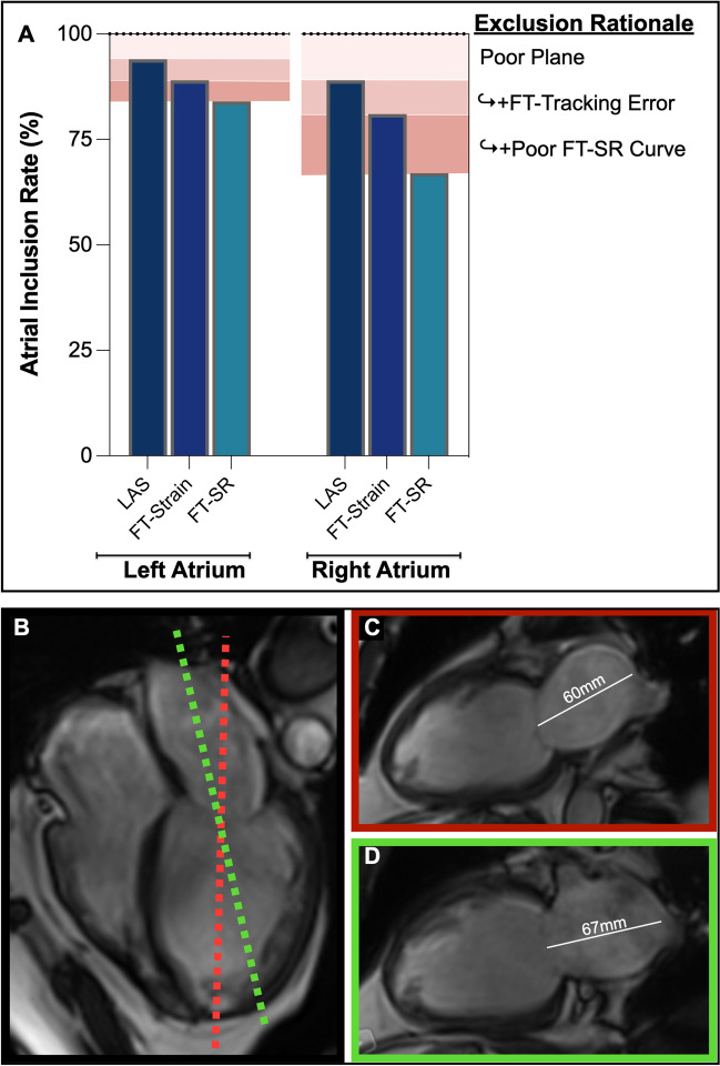
Atrial planes. The proportion of atrial images included in the analysis (A) indicates that while 6% of left atrial images and 11% of right atrial images were excluded for a poor plane position, all remaining images could be used for long-axis shortening (LAS) analysis, while additional images were excluded from feature tracking (FT) analysis due to tracking errors and poor strain rate (SR) curves. The lower panel from a patient with atrial fibrillation demonstrates that while the plane indicated in panel C (red) is the ideal location for ventricular analysis cutting perpendicular through the mitral valve and ventricular apex, this can lead to foreshortening of the left atrium (B), while panel D (green) depicts the true atrial length. However, image acquisition is typically localized off the ventricle leading to the proportion of datasets excluded due to the off-angle plane of the atria

