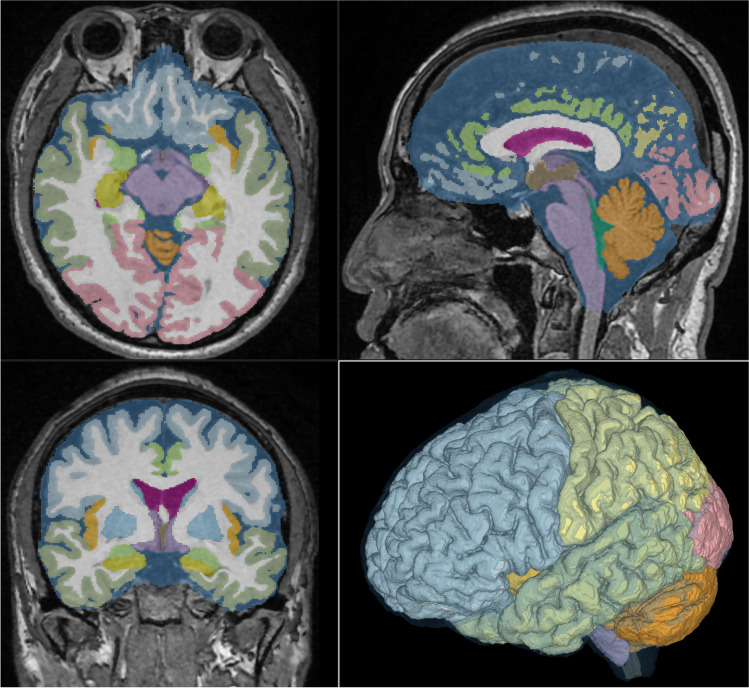
Fig. 2.
Screenshot of automatic segmentation using Brain Subregion Analysis application. This screenshot shows the result of a 48-year-old male healthy brain on the Brain Subregion Analysis application of the 3D volume analyzer SYNAPSE 3D workstation (FUJIFILM Corporation). By default, the axial view is displayed in the upper left, the sagittal view is in the upper right, the coronal view is in the lower left, and 3D is in the lower right

