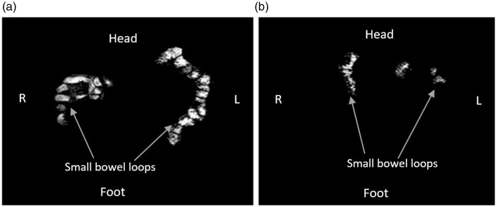
Fig. 4.
Coronal images of the abdomen in a single representative participant with the freely mobile water in the small bowel showing as bright white areas for F + ALG (a) and F-ALG (b), taken at 210 min after the commencement of feeding. More freely mobile water is evident with more bright white areas in A with F + ALG. (F + ALG, alginate-containing feed; F–ALG, alginate-free feed).

