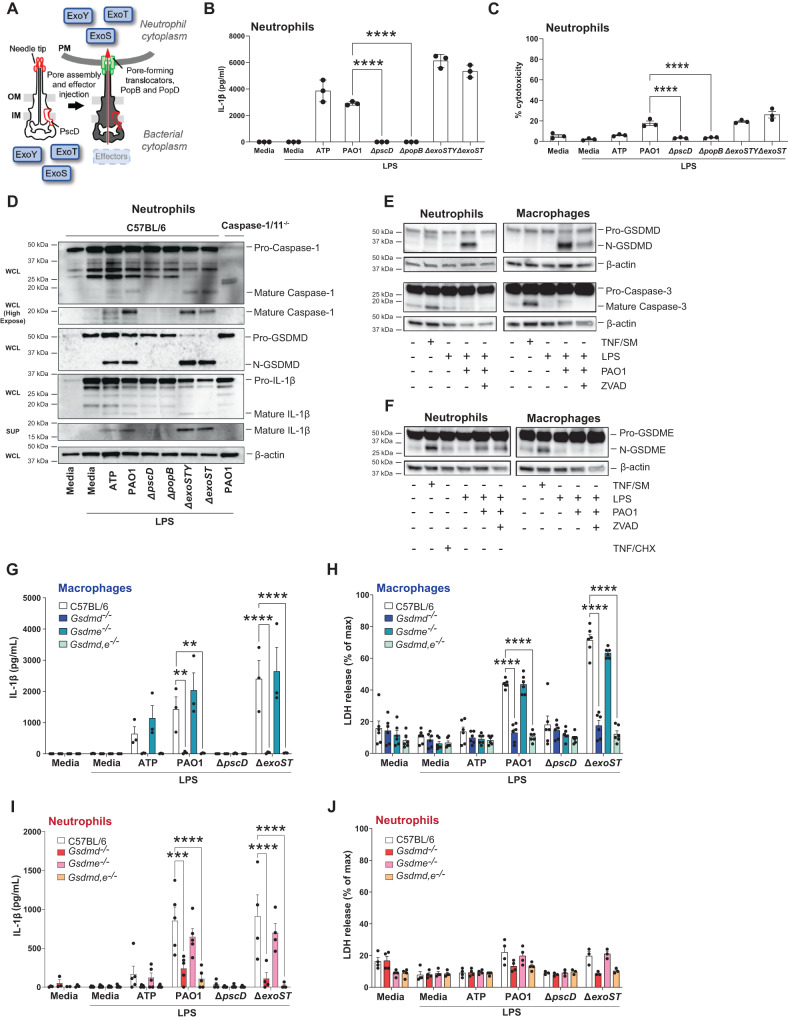
Fig. 1. The role of T3SS, GSDMD and GSDME in IL-1β secretion by P. aeruginosa infected neutrophils.
A P. aeruginosa T3SS is preassembled and spans the bacterial cell envelope (IM, inner membrane, OM, outer membrane and PG, peptidoglycan layer). Following cell contact, the pore-forming translocator proteins, PopB and PopD, assemble into a pore, which docks to the needle tip (PcrV, red). Effector secretion is triggered, resulting in export of effector proteins across the bacterial cell envelope and host cell plasma membrane (PM) into the cytosol of the targeted cell. P. aeruginosa strain PAO1 used in the current study expresses ExoS, ExoT, and ExoY, but not ExoU. B Quantification of IL-1β by ELISA, and C Cell death quantified by lactose dehydrogenase (LDH) activity in the supernatant of infected cells. D Caspase-1, GSDMD and IL-1β cleavage in whole cell lysates (WCL) and supernatants (SUP) from C57BL/6 and caspase-1/11-/- bone marrow neutrophils primed 3 h with LPS and incubated 1 h with ATP or 1 h with P. aeruginosa strain PAO1 or PAO1 mutants ∆pscD (no needle), ∆popB (no translocon), ∆exoSTY or ∆exoST. E, F Caspase-3, GSDMD and GSDME cleavage in lysates with supernatants from C57BL/6 bone marrow neutrophils and bone marrow derived macrophages primed with LPS and infected with PAO1 in the presence of ZVAD. TNF-α and SMAC mimetic (TNF/SM) induced caspase-3 / GSDME cleavage after 4 h (no LPS priming). G–J IL-1β secretion and LDH release from infected bone marrow-derived macrophages and bone marrow neutrophils from C57BL/6, Gsdmd-/- Gsdme -/- and Gsdmd/e-/- mice primed with LPS and incubated with PAO1 or indicated mutants. Western blots are representative of 3 repeat experiments. Each data point represents one independent experiment (3-4 biological replicates); error bars are mean + /- standard deviation (SD). Statistical significance was assessed by 1-way ANOVA followed by a Brown-Forsythe post-test (B, C), or using 2-way ANOVA followed by Tukey’s post-test (G-J). For all figures, ****p < 0.0001, ***p < 0.001; **<0.01.

