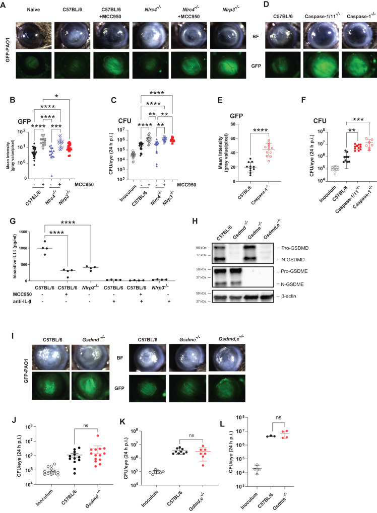
Fig. 4. The role of inflammasomes, caspase-1, GSDMD and GSDME in P. aeruginosa corneal infections.
A-C Corneas of MCC950 treated C57BL/6, Nlrc4-/-, and Nlrp3-/- mice were infected with 5×10⁴ GFP expressing PAO1. After 24 h, corneal opacification and total GFP bacteria were quantified by image analysis, and viable bacteria were measured by CFU. A representative images of infected corneas and GFP-PAO1. B Quantification of GFP in infected corneas. C CFU in infected corneas. D–F Corneal opacification, GFP-PAO1 and CFU in C57BL/6, caspase-1 and caspase-1/11-/- mice. G Bioactive IL-1β in infected corneas from C57BL/6, MCC950 treated mice, and Nlrp3-/- mice using the IL-1R reporter cells in the presence of neutralizing anti-IL-1β H–L PAO1 infected C57BL/6, gsdmd-/-, gsdme -/- and gsdmd / gsdme -/- corneas. H GSDMD and GSDME cleavage in infected corneas. I Representative corneas, and CFU (J–L) GSDMD and GSDME cleavage products were examined by western blot. Western blots are representative of 3 repeat experiments. Each data point represents a single infected cornea from 3 independent experiments with 4 infected corneas. Statistical significance was assessed by 1-way ANOVA followed by Kruskal-Wallis post-test for in vivo analysis. **** represents p < 0.0001, *** is p < 0.001; ** is <0.01, * is <0.05.

