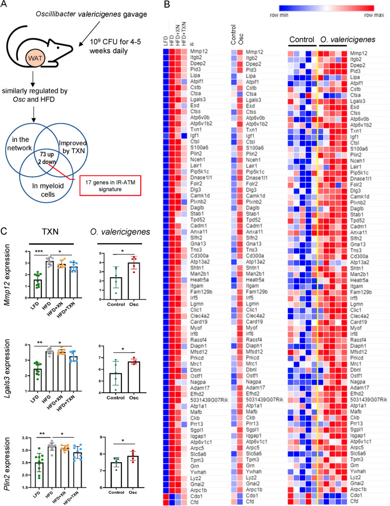
Fig. 5.
O. valericigenes treatment increases the expression of IR-ATM genes in mouse white adipose tissue that are improved by TXN treatment. A Outline of O. valericigenes experiment and subsequent gene expression analysis from WAT (n = 5 per group). The Venn diagram shows 75 genes from myeloid cells overlapping between similar effects of HFD and O. valericigenes with opposite effects of TXN. WAT genes from the network in Fig. 1D. B Expression of adipose tissue genes increased by HFD and decreased by TXN treatment in mice (left panel) vs the same adipose tissue genes following O. valericigenes supplementation of mice (right panel). The middle panel shows the mean of the samples in the right panel. Colors represent the quantile normalized log2(CPM+1) mean of each group, relative to the other groups in the row (e.g., the darkest blue color indicates the lowest mean while dark red indicates the highest). C Comparison of in vivo adipose tissue gene expression in two sets of experiments. One experiment used varying diets (low fat diet and high fat diet) and interventions (XN and TXN treatment) while the other used only a normal chow diet with or without O. valericigenes supplementation. Shown are examples of adipose tissue genes (in the IR-ATM signature) decreased by TXN treatment but induced by O. valericigenes (Osc) supplementation in vivo. Values are in normalized counts per million. *Mann–Whitney p-value < 0.05; **p-value < 0.01; ***p-value < 0.001

