Fig. 6.
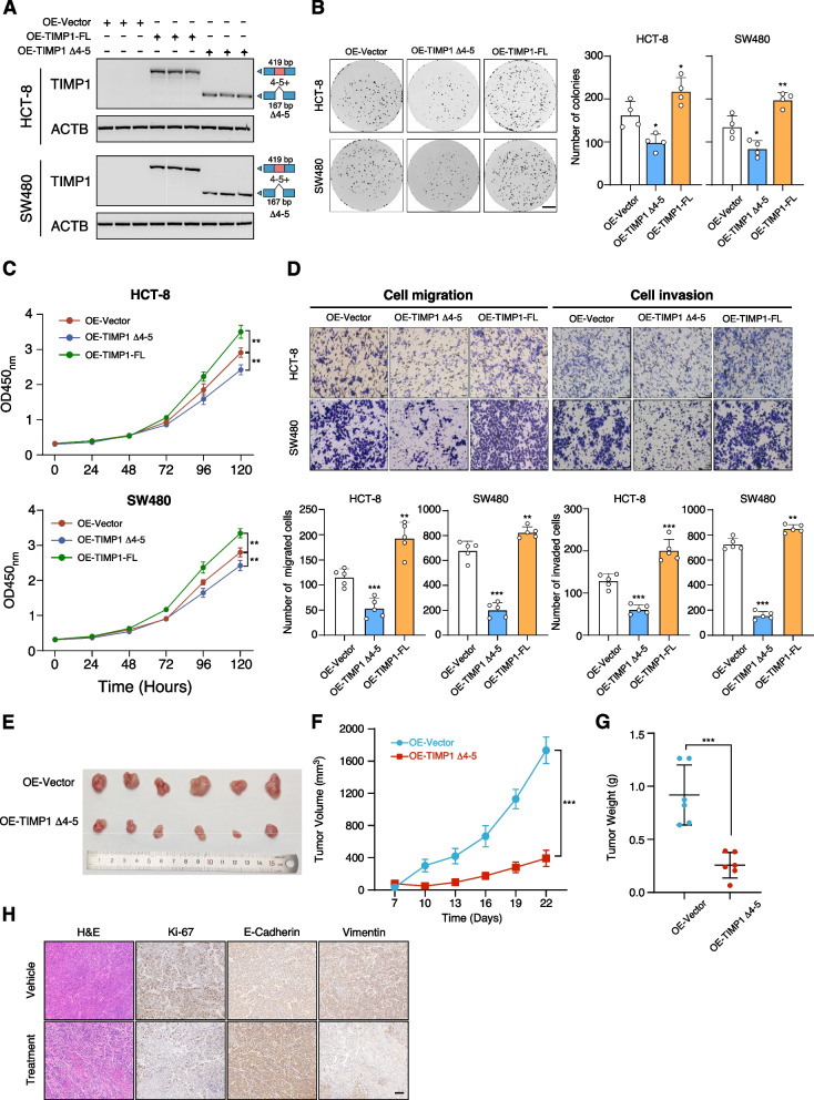
TIMP1 Δ4-5 and TIMP-FL have antithetical functions in CRC carcinogenesis. A Exogenous overexpression of TIMP1 Δ4-5 and TIMP-FL in TIMP1-KD HCT-8 and SW480 cells. B, C Colony formation (B) and MTT (C) assay of exogenous overexpression of TIMP1 Δ4-5 and TIMP-FL in TIMP1-KO HCT-8 and SW480 cells. P values were calculated by two-sided Student’s t test, * P < 0.05, ** P < 0.01. D Tumor cell migration and invasion assay of exogenous overexpression of TIMP1 Δ4-5 and TIMP-FL in TIMP1-KO HCT-8 and SW480 cells. P values were calculated by two-sided Student’s t test, ** P < 0.01, *** P < 0.001. The migrated or invaded cells were quantified by counting in five fields. Scale bar, 100 μm. E Xenograft mouse model established using SW480 cells was stably infected with lentivirus-based TIMP1 Δ4-5 or empty vector in BALB/c nude mice (n = 6 mice per group). In vivo generated tumors are depicted. F, G Analysis of tumor growth (F) and weight (G) in the xenograft mouse model. Data are presented as mean ± SEM of n = 6 mice per group. Two-way ANOVA and one-way ANOVA followed by Tukey test. H Representative H&E and IHC images of randomly selected tumors were shown. Scale bar, 100 µm

