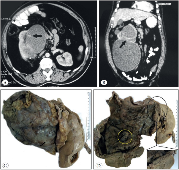
Figure 1.
CECT: A) Axial: cystic mass arising from upper pole with solid enhancing nodular projection (arrow). B) Coronal reconstruction: thick internal septae (arrow). C) Total nephrectomy specimen showing large cystic tumor. D) Well-defined renal interface (black circle), extrarenal extension with cortical cyst (arrow) and thickened mural nodule (yellow circle)

