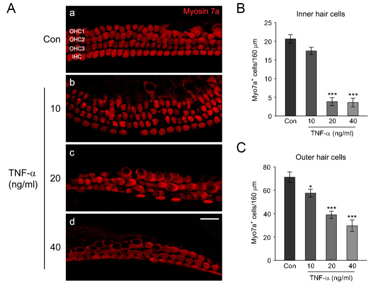
Fig 5. Effects of TNF-α on the cochlear hair cells in middle-turn cochlear explants.
(A) The representative confocal images show the middle-turn cochlear explants treated with a culture medium alone (control) or a medium containing different concentrations of TNF-α (10, 20, and 40 ng/ml). After treatment, explants were fixed, permeabilized, and stained with a polyclonal myosin 7a antibody as a hair cell marker. Scale bars = 20 mm, Original magnification = 400 ×. (B, C) Quantification of myosin 7a positive Inner hair cells (IHCs) and Outer hair cells (OHCs) per 160 mm in the middle-turn cochlear explants, respectively. Data are expressed as mean ± SE of the number of IHCs or OHCs (n = 3 different explants per group); * P < 0.05, *** P < 0.001, *,***; compared with untreated control.

