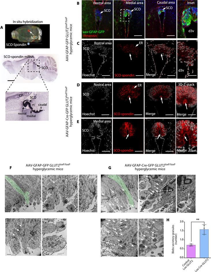
Fig 3. When the CSF glucose concentration is increased, the release of secretory granules by SCO cells is decreased in GLUT2loxP/loxP mice after genetic inactivation of GLUT2.
(A) 3D brain imaging and sagittal sections after in situ hybridization showing SCO-spondin expression restricted to the commissural area epithelia of the third ventricle in the adult mouse brain. SCO-spondin is shown in red in the 3D brain image, and in blue, in the sagittal sections. Scale bar: 2 mm. Image credit: Allen Institute. (B) Immunohistochemical staining of vimentin in frontal brain sections and analysis of AAV-GFAP-GFP expression in GLUT2loxp/loxp mice. Scale bar: 400 μm. (C) SCO-spondin (red) and Hoechst (white; nuclei) staining in AAV-GFAP-GFP–injected hyperglycemic GLUT2loxp/loxp mice. Secretory SCO-spondin was detected in ER cisternae; however, secretory apical granules were not detected (arrow and inset). Scale bar: 200 μm. (D and E) SCO-Spondin (red) and Hoechst (white; nuclei) staining in AAV-GFAP-Cre-GFP–injected hyperglycemic GLUT2loxp/loxp mice. Secretory SCO-spondin was detected in ER cisternae and secretory apical granules (arrow and inset). Scale bar: 200 μm. (F and G) TEM analysis of AAV-GFAP-GFP–injected hyperglycemic GLUT2loxp/loxp and AAV-GFAP-Cre-GFP–injected hyperglycemic GLUT2loxp/loxp mice. Elongated SCO cells (green cells) were visualized at low magnification, and apical blebs were visualized at higher magnification. The secretory granules are indicated by white arrows. Scale bar: lower magnification, 2 μm; higher magnification, 0.5 μm. (H) Quantification of the number of SCO cell secretory granules in AAV-GFAP-Cre-GFP–injected hyperglycemic GLUT2loxp/loxp mice and AAV-GFAP-GFP–injected hyperglycemic GLUT2loxp/loxp mice. The graph shows data from 3 biologically independent samples. The error bars represent the SD; **P < 0.01 (two-tailed Student t test). Data used to generate graph can be found in S1 Data. CSF, cerebrospinal fluid; d3v, dorsal third ventricle; ER, endoplasmic reticulum; GLUT2, glucose transporter 2; MT, microtubule; MV, microvilli; SCO, subcommissural organ.

