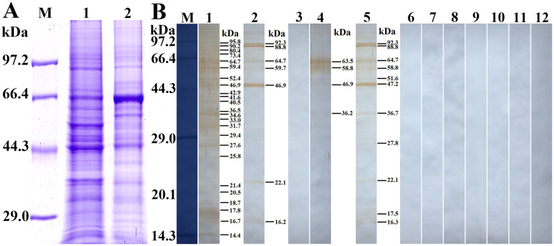
Fig 12. Far-Western analysis of rTsSPc binding to Caco-2 cell proteins.
The Caco-2 cell proteins were first analyzed by SDS-PAGE, subsequently the rTsSPc binding with Caco-2 cell protein was detected on Far-Western analysis. A: SDS-PAGE analysis of Caco-2 cell proteins. Lane M: protein marker; Lane 1: Caco-2 cell lysates; Lane 2: C2C12 cell lysates as a cell negative control. B: Far-Western analysis of rTsSPc binding to Caco-2 cell proteins. Caco-2 cell proteins were first incubated with rTsSPc (Lanes 1–3), IIL ES antigens (Lanes 4–6) or BSA (Lanes 7–9), and then probed by anti-rTsSPc serum (Lanes 1, 4 and 7), infection serum (Lanes 2, 5 and 8), and pre-immune serum (Lanes 3, 6 and 9); The C2C12 protein (Lanes 10–12) was first incubated with rTsSPc, and subsequently probed by anti-rTsSPc serum (Lane 10), infection serum (Lane 11) or pre-immune serum (Lane 12). There was no binding between rTsSPc and C2C12 protein.

