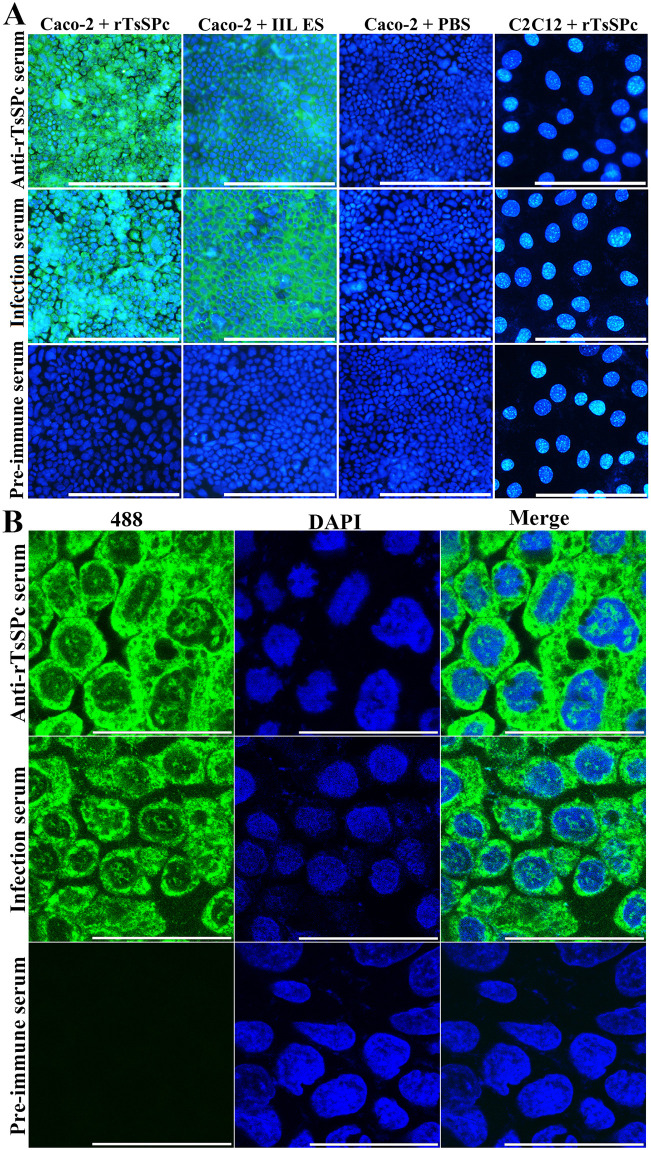
Fig 13. Binding of rTsSPc and Caco-2 cells by IIFT and confocal microscopy.
A: IIFT analysis of binding between rTsSPc and Caco-2 cells (200×). Caco-2 cells were first incubated with rTsSPc, IIL ES antigens or PBS for 2 h at 37 °C. C2C12 cells were also incubated with rTsSPc for 2 h at 37 °C. After washes, the cells were probed by anti-rTsSPc serum, infection serum or pre-immune serum, subsequently colored with goat anti-mouse IgG-488 conjugate. 4’,6-diamidino-2-phenylindole (DAPI) dyed cell nuclei in blue. Scale bars: 200 μm. B: Cellular localization of rTsSPc binding to Caco-2 by confocal microscopy (1000×). The binding sites were localized in cell membrane and cytoplasm Abbreviations: 488: Alexa Fluor 488; DAPI, 4’,6-diamidino-2-phenylindole. Scale bars: 40 μm.

