FIGURE 5.
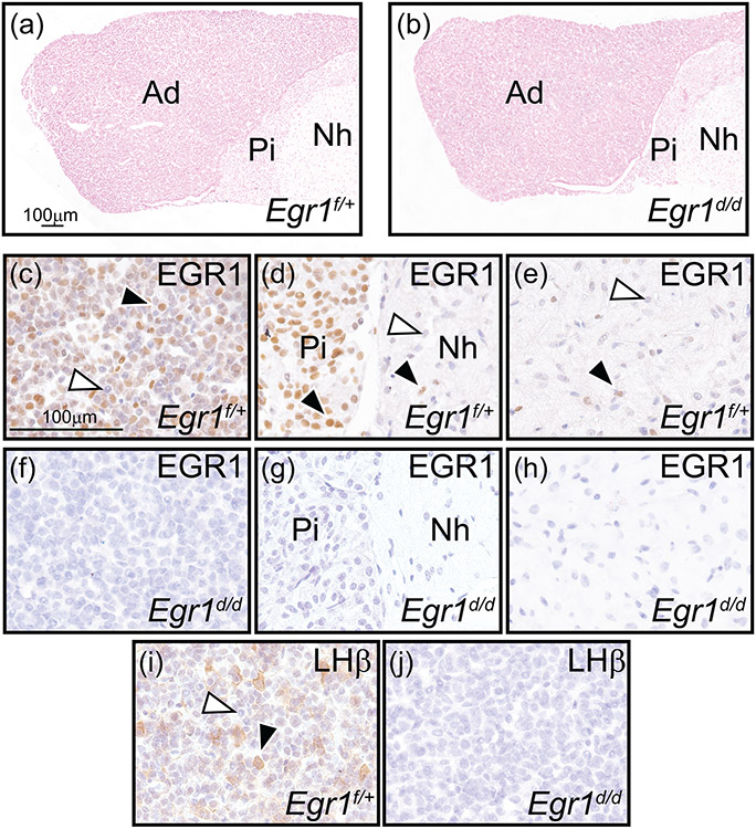
Absence of EGR1 and LH-β subunit expression in the female Egr1d/d pituitary gland. (a) and (b) show representative nuclear-fast red stained sections of Egr1f/+ and Egr1d/d pituitary gland tissue respectively; representative of four adult (nine weeks old) mice per genotype. Pituitary gland size and histomorphology were equivalent for both genotypes (adenohypophysis (or the anterior pituitary gland or pars distalis), pars intermedia (or intermediate gland), and neurohypophysis (or posterior pituitary gland or pars nervosa) are indicated by Ad, Pi, and Nh respectively). Scale bar in (a) also applies to (b). (c-e) Immunohistochemical analysis of EGR1 expression in the Ad, Pi, and Nh of the Egr1f/+ pituitary gland respectively. Note in (c) the presence of numerous EGR1 positive cells in the Ad (black arrowhead); a subgroup of Ad cells is EGR1 negative (white arrowhead). (d) The majority of cells in the Pi region is immunopositive for EGR1 (black arrowhead), with few cells EGR1 positive in the Nh region ((d) and (e) (black arrowhead)); EGR1 negative cells in the Nh are indicated by a white arrowhead ((d) and (e)). (f-h) Expression of EGR1 is absent in all regions of the Egr1d/d pituitary gland. (i) Immunohistochemical detection of the LH β subunit in the Ad region of the Egr1f/+ pituitary gland. Note a subset of Ad cells is positive for LH β expression (black arrowhead); the white arrowhead indicates an Ad cell that is LH β negative. (j) Expression of the LH β subunit is absent in the Ad region of the Egr1d/d pituitary gland. Scale bar in (c) applies to (d-j).

