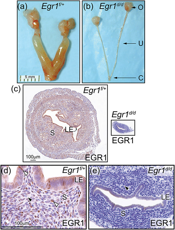
Figure 6.
Abrogation of EGR1 results in a hypoplastic uterus in the Egr1d/d mouse. (a) and (b) show the gross morphology of the Egr1f/+ and Egr1d/d female reproductive tract respectively; representative of five adult mice per genotype. Compared with the Egr1f/+ uterus (a), note the thin Egr1d/d uterine horn (b); ovary, uterus, and cervix are denoted by O, U, and C respectively. Scale bar in (a) applies to (b). (c) Transverse section of the mid-region of the Egr1f/+ and Egr1d/d uterine horn (left and right panels respectively) is shown; luminal epithelium and stroma are indicated by LE and S respectively. Both tissue sections were immunohistochemically stained for EGR1 expression. Note EGR1 expression in S and LE compartments of the Egr1f/+ uterus whereas EGR1 expression is not detected in the Egr1d/d uterus. Scale bar in the left panel applies to right panel. (d) and (e) are higher magnification images of the micrographs shown in the left and right panels in (c) respectively. (d) Expression of EGR1 is detected in subsets of cells in the S and LE compartment of the Egr1f/+ uterus (white arrowhead). (e) Expression of EGR1 is absent in the Egr1d/d uterus (black arrowhead). Scale bar in (d) applies to (e).

