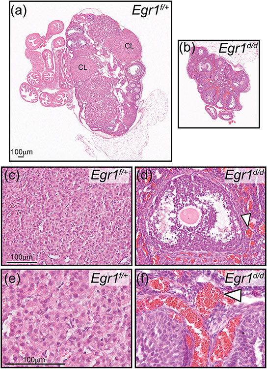
Figure 7.
Absence of corpora lutea in the atrophied Egr1d/d ovary. (a) Hematoxylin and eosin stained section of the Egr1f/+ ovary; a corpus luteum is indicated by CL. (b) Similarly stained Egr1d/d ovarian tissue section; note the absence of CLs. Also note the diminished size of the Egr1d/d ovary compared with the Egr1f/+ ovary shown in (a). Scale bar in (a) applies to (b). The histological data are representative of five adult mice per genotype. (c) Field of view shows luteal cells within the CL of an Egr1f/+ ovary. (d) Image shows an antral follicle within the Egr1d/d ovary; note the juxtaposed hemorrhagic area indicated by the white arrowhead. Scale bar in (c) applies to (d). (e) and (f) represent higher magnification images of regions in (c) and (d) respectively. Again, note the hemorrhagic areas in the Egr1d/d ovary (white arrowhead). Scale bar in (e) applies to (f).

