Figure 8.
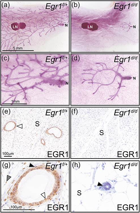
A prepubescent mammary gland phenotype in the adult Egr1d/d mouse. (a) Whole mount of an inguinal mammary gland from an adult Egr1f/+ mouse, note the extensive epithelial ductal branching from the nipple (N) to the periphery of the fat pad. The lymph node is indicated by LN, which is used as a structural reference point in the inguinal gland. (b) Absence of epithelial ductal elongation and dichotomous branching in the mammary of the adult Egr1d/d mouse. Note that the majority of the Egr1d/d mammary gland fat pad is devoid of the epithelial compartment. Mammary gland results are representative of five adult mice per genotype. Scale bar in (a) applies to (b). (c) and (d) represent higher magnification images shown in (a) and (b) respectively; scale bar in (c) applies to (d). (e) Immunohistochemical detection of EGR1 expression in a section derived from Egr1f/+ mammary gland tissue. Immunopositivity in the mammary epithelium is indicated by white arrowhead; mammary stroma is denoted as S. (f) Immunopositivity for EGR1 is absent in the mammary gland of the Egr1d/d mouse. As indicated in panel (b), the majority of the mammary gland is devoid of the epithelial compartment in the Egr1d/d mouse; scale bar in (e) applies to (f). (g) Higher magnification image clearly showing EGR1 expression in the basal epithelium (black arrowhead), a subset of luminal epithelial cells (white arrowhead), and stromal cells (gray arrowhead). (h) Immunopositivity for EGR1 is not detected in cells of vestigial epithelial ducts that are localized to the nipple region of the adult Egr1d/d mammary gland; the stromal compartment is also negative for EGR1 immunopositivity. Scale bar in (g) applies to (h).

