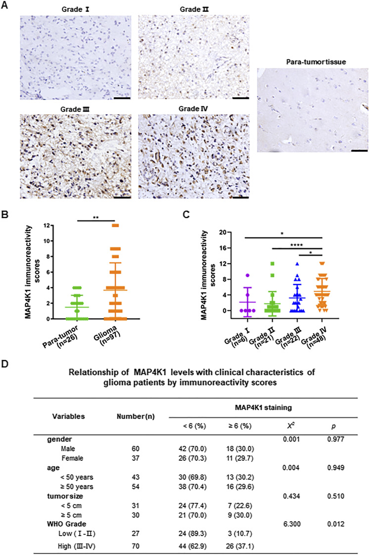
Figure 1. MAP4K1 expression is elevated in human high-grade gliomas.
Human glioma (n = 97) and para-tumor tissues (n = 26) were stained with standard MAP4K1 immunohistochemistry (IHC). (A) Representative IHC images of MAP4K1 expression in human glioma tissues. Scale bar, 50 μm. (B) Quantification of MAP4K1 immunoreactivity scores in human para-tumor and glioma tissues. Data are represented as single data points and mean ± SD. **P < 0.01 (Mann‒Whitney U test). (C) Quantification of MAP4K1 immunoreactivity scores in grades I–IV of gliomas (n = 6, 21, 22, 48). Data are represented as single data points and mean ± SD. *P < 0.05, ****P < 0.0001 (one-way ANOVA). (D) Correlation analysis of pathological characteristics of glioma patients with MAP4K1 expression according to MAP4K1 IHC scores. Data were analyzed by the Mann–Whitney U test and χ2 test.

