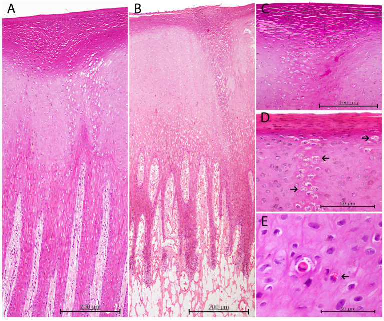
Figure 3.
Histopathological findings in CePV-1 positive skin lesions from five cases. (A) Lesion A1 from case 7. Focal marked hyperkeratosis showing two focal columns of ballooning degeneration affecting apical areas of rete ridges and the epidermal transitional zone between both stratums corneum and spinosum. H and E, ×10. (B) Lesion A1 from case 16. Focal zone of moderate ballooning degeneration affecting both stratum corneum and spinosum. Marked hyperkeratosis just above the line of vacuolated keratinocytes is observed. Marked multifocal congestion in the dermal papillae. H and E, ×10. (C) Lesion A6 from case 11. Marked focal hyperkeratosis. Beneath this affected area, a moderate focal ballooning degeneration in the stratum spinosum is appreciated. H and E, ×20. (D) Lesion A1 from case 30. ICIBs detected in a column-like group of vacuolized keratinocytes (arrows). Right above, mild hyperkeratosis with associated slightly hyperpigmented keratinocytes. HE, ×40. (E) Lesion A1 from case 31. Acidophilic apoptotic keratinocyte with small amphophilic ICIBs. Multiple irregular sized ICIBs in a vacuolated keratinocyte (arrow). H and E, ×40.

