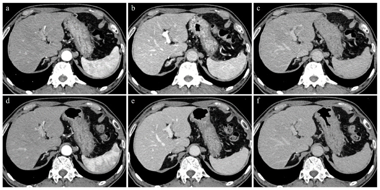
Figure 1.
A 74-year-old man with 62.3kg body weight. (a–c) Initial CT images obtained with 70 kVp protocol during AP (a), PVP (b), and EP (c). (d–f) Follow-up CT images (9 months later) obtained with blended DE protocol during AP (d), PVP (e), and EP (f). All CT images were reconstructed by IR methods. Contrast enhancement of the hepatic parenchyma, portal vein, and hepatic vein in PVP and EP were better in 70 kVp than in blended DE protocol.

