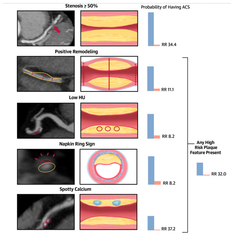
Figure 2.
Central illustration. Significant stenosis and high-risk coronary plaque features and their association with the probability of ACS during index hospitalization. Stenosis ≥ 50%: severe stenosis of the mid-left anterior descending coronary artery (red arrow). Positive remodeling: noncalcified plaque with positive remodeling in the distal right coronary artery. The 2 dotted red lines demonstrate the vessel diameters at the proximal and distal references (both 1.8 mm), and the solid red line demonstrates the maximal vessel diameter in the mid portion of the plaque (2.7 mm). The remodeling index is 1.5. Low Hounsfield units (HU) plaque: partially calcified plaque in the mid-right coronary artery with low <30 HU plaque. The red circles demonstrate the 3 regions of interest, with mean computed tomography (CT) numbers of 22 HU, 19 HU, and 20 HU. Napkin-ring sign: napkin-ring sign plaque in the mid-left anterior descending coronary artery. Schematic cross-sectional view of the napkin-ring sign. The red line demonstrates the central low HU area of the plaque adjacent to the lumen (yellow ellipse) surrounded by a peripheral rim of the higher CT attenuation (red arrows). Spotty calcium: partially calcified plaque in the mid-right coronary artery with spotty calcification (diameter < 3 mm in all directions; red circles). Patients with significant CAD showing ≥50% stenosis were at a significantly higher risk of experiencing an ACS during their hospitalization, being 34 times more likely compared to those without significant CAD. Furthermore, the presence of high-risk plaque, including positive remodeling (RR 11.1), low Hounsfield unit (HU) or napkin-ring sign (NRS) features (both with a RR of 8.2), spotty calcium (RR 37.2), or at least 1 high-risk plaque feature (RR 32), was associated with an elevated relative risk of ACS. ACS = acute coronary syndromes; RR = relative risk. Source: Puchner SB, High-Risk Plaque Detected on Coronary CT Angiography Predicts Acute Coronary Syndromes Independent of Significant Stenosis in Acute Chest Pain: Results From the ROMICAT-II Trial, Volume 64, Issue 7, 19 August 2014, Pages 684–692 [11].

