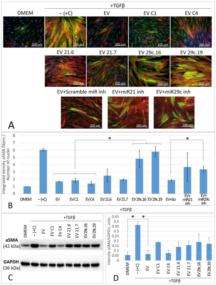
Figure 4.
Content analysis of the myofibroblast marker alpha smooth muscle actin (aSMA) in the cultures of fibroblasts treated with TGFb combined with EV fraction obtained from native or CRISPR/Cas9-modified ASC52telo cells. (A)—Immunocytochemical staining: alpha smooth muscle actin—green, total actin (phalloidin)—red, DAPI—blue. (B)—Relative level of cell culture staining intensity (stain for the marker protein of myofibroblasts aSMA), *—p < 0.05, n ≥ 4. (C)—Western blot analysis. (D)—Quantitative analysis of Western blot results, normalized for GAPDH, *—p < 0.05, n ≥ 2.

