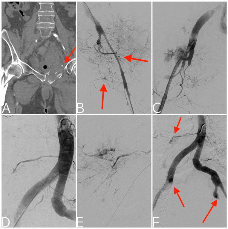
Figure 3.
Pedestrian versus motorcycle accident. CT angiography with multiplanar reconstruction in the coronal plane depicting right femoral neck and left superior pubic ramus fractures with active contrast extravasation (arrow) (A). Digital subtraction angiography documenting severe vasospasm of the left internal iliac artery, along with active bleeding from both anterior and posterior divisions (B). Subsequent state of hemodynamic instability with consequent proximal prophylactic embolization using a gelatin sponge. Contralateral digital subtraction angiography depicting vasospasm of the right internal iliac artery together with severe arterial injury of the posterior division (C). This is followed by embolization using an EVOH copolymer. Aortogram showing right fifth lumbar artery bleeding (D), confirmed with superselective catheterization (E) and successfully managed with coils embolization. Final aortogram depicting the bilateral proximal embolization of both internal iliac arteries and right fifth lumbar artery embolization (arrows) (F).

