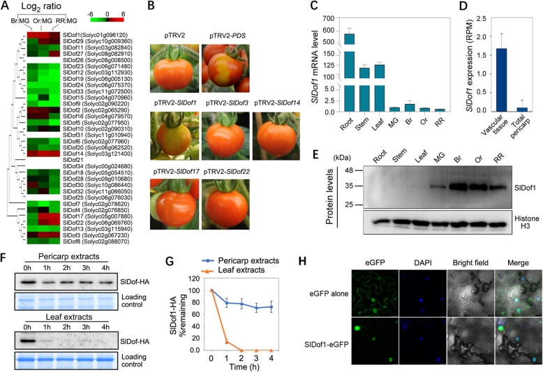
Fig. 1.
Virus-induced gene silencing (VIGS) screening and expression analysis reveal the involvement of SlDof1 in tomato fruit ripening. (A) Phylogenetic analysis of tomato Dof genes and expression profiles during fruit ripening, as determined by quantitative RT-PCR. The phylogenetic tree was produced using the neighbor-joining method in MEGA version 6.0 with bootstrapping analysis (1000 replicates). The ACTIN gene was utilized as an internal control. The ripening stages include mature green (MG), breaker (Br), orange (Or), and red ripe (RR). Expression ratios were plotted in a heat map on a log2 scale, using the MG stage as the denominator. Each row in the color heat map represents a single Dof gene, and the gene identifiers (Solyc numbers) are shown. Empty box indicates no expression in fruit. Data from biologically repeated samples are averaged. (B) VIGS assay revealing the involvement of SlDof1 in fruit ripening. Images show the ripe fruit of plants infected with vectors containing no insert (pTRV2; negative control), a specific fragment of phytoene desaturase (PDS) (pTRV2-PDS; positive control), or a specific Dof sequence. (C) Expression of the SlDof1 gene in roots, stems, leaves, and pericarps of fruit at different ripening stages, as determined by quantitative RT-PCR. Values are means ± SD of three independent experiments. (D) Expression of SlDof1 in the vascular tissue of the pericarp and the total pericarp based on the Tomato Expression Atlas database (http://tea.solgenomics.net/). RPM, reads per million mapped reads. (E) Western blot analysis of SlDof1 protein in the roots, stems, leaves, and pericarps of fruit. An anti-histone H3 immunoblot was used as a protein loading control. (F) Cell-free degradation assay of SlDof1. The recombinant SlDof1-HA protein was purified and incubated in extract from tomato pericarps or leaves. The protein levels at different time intervals were measured by immunoblotting using an anti-HA antibody. (G) Quantification of protein levels in (F) by ImageJ. (H) Subcellular localization of SlDof1. Nicotiana benthamiana leaves transiently expressing eGFP alone (control) and SlDof1-eGFP were observed under a Leica confocal microscope. The fluorescent dye 4′,6-diamidino-2-phenylindole (DAPI) was used for nuclear staining. Scale bars, 25 μm

