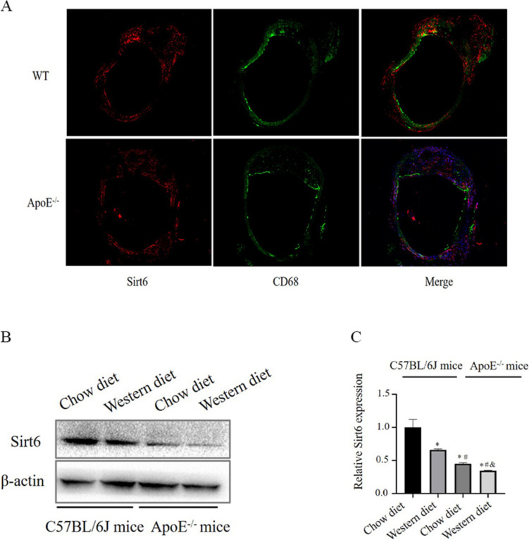
Fig. 1.
Sirt6 was observed in macrophage-derived foam cells. A WT mice and ApoE−/− mice (n = 6) with atherosclerotic plaques were given a western diet (WD) for 16 weeks, stained (red) for Sirt6 and CD68 (green), and co-localised (yellow merging); DAPI staining was performed on the nuclei (blue). B Immunoblots demonstrating the expression of Sirt6 in the aorta obtained from wild-type C57BL/6J mice and ApoE−/− mice given a chow diet and western diet (n = 10). C Quantification of Sirt6 expression in each group (n = 10) (One way ANOVA showed that there was statistical significance among groups. *P < 0.05 versus (vs) WT mice [n = 10]; #P < 0.05 vs ApoE−/− mice [n = 10]; $P < 0.05 vs ApoE−/−: Sirt6−/− mice [n = 10]

