Fig. 2.
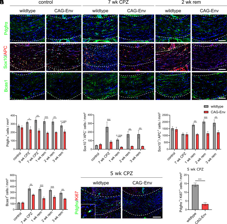
Transgenic HERV-W ENV expression affects oligodendroglial differentiation. (A) Representative immunohistochemical images of Pdgfrα-, Sox10/APC- and Bcas1- expression in unchallenged animals (wt and CAG-Env mice), after 7 wk of CPZ treatment and after 2 wk of CPZ withdrawal (2 wk rem). (B) Quantification of Pdgfrα-positive cell densities in the corpus callosum of control vs. CPZ-treated animals. (C) Quantification of Sox10-positive, APC-negative cell densities in the corpus callosum of control vs. CPZ-treated animals. (D) Quantification of Sox10/APC double-positive maturing oligodendroglial cell densities in the corpus callosum of control vs. CPZ-treated mice. (E) Quantification of Bcas1-positive myelinating oligodendrocyte densities in the corpus callosum of control vs. CPZ-treated animals. (F) Representative immunohistochemical pictures of Pdgfrα/Ki67-coexpressing cells in the corpus callosum of wt vs. CAG-Env mice at 5 wk of CPZ treatment. (G) Quantification of Ki67-positive proliferating OPCs in wt vs. CAG-Env corpus callosum tissues after 5 wk of CPZ diet. Data are presented as mean values (n = 6) ± SEM. Significance of Ki67-positive OPCs was analyzed by Student’s unpaired t test, whereas all other significances were accessed by 2-way ANOVA followed by Sidak’s post hoc test (95% CI) at *P < 0.05, **P < 0.01, ***P < 0.001. Dashed lines indicate the area of corpus callosum. (Scale bar: 100 µm.)

