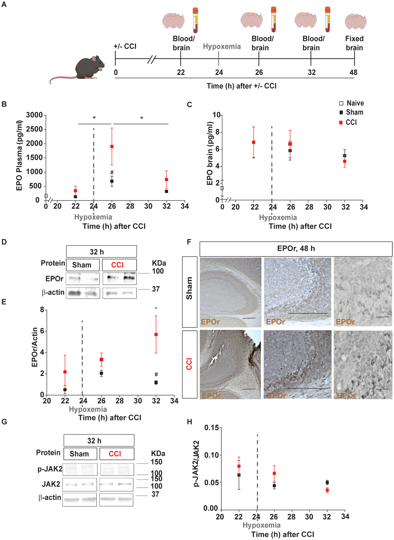
Fig. 1.

Delayed hypoxemia increased EPO and EPOr synthesis in the blood and brain, respectively, following TBI. A, Experimental design: brain and blood collection 22h, 24h, 26h, 32h and 48h after injury inducing 1h of hypoxemia at 24h post-injury. B, Endogenous mouse EPO in plasma over time analyzed by ELISA after TBI with delayed hypoxemia. C, Endogenous mouse EPO in the brain over time analyzed by ELISA after TBI with delayed hypoxemia. D, Representative western blot of the hippocampus membrane fraction probed for EPOr and (E) densitometric analysis. F, Representative image of EPOr-positive cells in CA3 region of injured hippocampi. G, Representative western blot of the hippocampus cytoplasm fraction probed for pJAK2 and (H) densitometric analysis. β-actin was used as a loading control. For western blot images, 2 samples per condition always from the same gel, using the same two samples throughout the figure. Two-way (Time and hypoxemia) ANOVA were used to determine statistical differences for hypoxemia effect followed by Tukey multiple comparison post-hoc test were used to determine statistical differences, n=6–7 mice per group. Time F(5,57) = 3.424 p = 0.009. Hypoxemia F(1,57) = 10.53 p < 0.0020. Scale bar: 20 μm. Abbreviations: CCI, controlled cortical impact; EPO, erythropoietin, EPOr, erythropoietin receptor.
