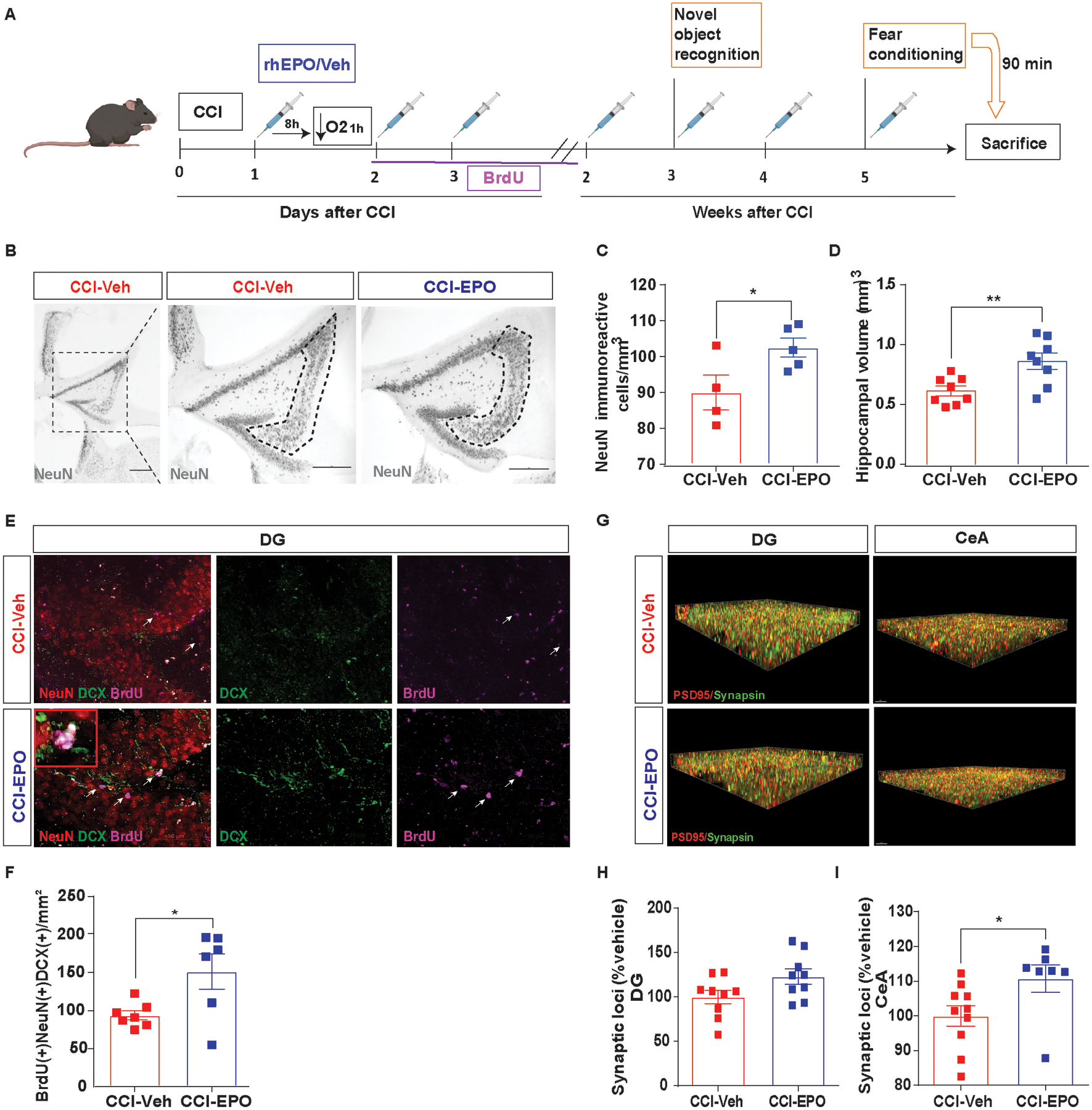
Fig. 3.

Ongoing rhEPO administration induced neuroprotection, increased neurogenesis in hippocampus and, increased synaptic density in amygdala. A, Experimental design. B, Representative image of NeuN+ cells in CA3 region of injured hippocampi (indicated by the dotted line) and (C) stereological quantification. D, Hippocampal volume quantification. E, Representative immunofluorescence image of the SGZ in the DG of the hippocampus labeled with NeuN (red), DCX (green) and BrdU (magenta) with a zoomed in insert. White arrows indicate BrdU-positives cells. F Summation of total neuronal lineage cells per area of hippocampi. G, Puncta detection of PSD95 (red) synapsin (green) images in DG (left) and CeA (right). H, Quantification of synaptic loci (% vehicle) in DG and (I) CeA. Unpaired t-test *p< 0.05, **p< 0.01, n=4–8 mice per group. Scale bar: 50 μm. Abbreviations: CCI, controlled cortical impact; rhEPO, recombinant human erythropoietin; Veh, vehicle; SGZ, subgranular zone; DG, dentate gyrus; DCX, doublecortin; BrdU, 5-bromo-2’-deoxyuridine. CeA, central amygdala.
