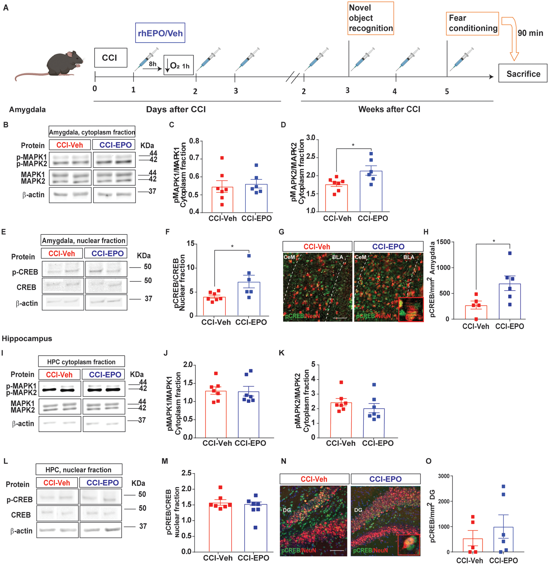
Fig. 4.

MAPK/CREB signaling pathway activation after rhEPO treatment in amygdala but not in hippocampus. A, Experimental design. B, Representative western blot of the amygdalar cytoplasm fraction probed for total and phosphorylated MAPK1 and 2. C, Densitometric analysis of pMAPK1 expression normalized by total MAPK1. D, Densitometric analysis of pMAPK2 expression normalized by total MAPK2. E, Representative western blot of the amygdalar nuclear fraction probed for total and phospho-CREB. F, Densitometric analysis of pCREB expression normalized by total CREB. β-actin was used as a loading control. G, Representative immunofluorescence image of the BLA labeled with pCREB (green) and NeuN (red) with a zoomed in insert. H, Quantification of pCREB density expression in neurons in the BLA. I, Representative western blot of the hippocampal cytoplasm fraction probed for total and phosphorylated MAPK1 and 2. J, Densitometric analysis of pMAPK1 expression normalized by total MAPK1. K, Densitometric analysis of pMAPK2 expression normalized by total MAPK2. L, Representative western blot of the hippocampal nuclear fraction probed for total and phospho-CREB. M, Densitometric analysis of pCREB expression normalized by total CREB. β-actin was used as a loading control. For western blot images, 2 samples per condition always from the same gel, using the same two samples throughout the figure. N, Representative immunofluorescence image of the BLA labeled with pCREB (green) and NeuN (red) with a zoomed in insert. O, Quantification of pCREB density expression in neurons in the BLA. Mean values are plotted ± SEM, One-way ANOVA followed by Tukey multiple comparison post hoc test were used to determine statistical differences; *p<0.05, n=5–7 mice per group. Mean values are plotted ± SEM, unpaired t-test *p<0.05, n=5–6 mice per group. Scale bar: 50 μm. Abbreviations: CCI, controlled cortical impact; rhEPO, recombinant human erythropoietin; Veh, vehicle; pCREB, phosphorylated cAMP-response element binding protein; pMAPK, phosphorylated mitogen-activated protein kinase; BLA, basolateral amygdala; CeA, central amygdala.
